Mai Mohammad Elgendy, Somaya Abdel-Gawad Madkour, Fatma Mohamed Magdi Badr Eldine, Doaa Mokhtar Emara, MennattAllah Hassan Attia
{"title":"Utility of the morphological scoring of costal cartilage ossification in age estimation of adult Egyptians using multidetector computed tomography.","authors":"Mai Mohammad Elgendy, Somaya Abdel-Gawad Madkour, Fatma Mohamed Magdi Badr Eldine, Doaa Mokhtar Emara, MennattAllah Hassan Attia","doi":"10.1093/fsr/owae061","DOIUrl":null,"url":null,"abstract":"<p><p>Age estimation of adults is a challenging procedure in forensic practice. Inspired by the previous work by Chinese scholars, we established population-specific age estimation models from the osseous and calcified projections (OCPs) of costal cartilages, using three-dimensional volume-rendering technique. A total of 168 clinical CT scans (2 mm slice thickness) were used to develop the sex-specific age prediction models from a sample of Egyptians, comprising 70 females and 98 males, with documented ages between 12 and 85 years. The sample was also used for validating the Chinese model. We reported the differences between the predictive accuracy of the Egyptian (population specific) and Chinese (non-population specific) models. The most accurate age estimation model was stepwise linear regression with standard error of estimates of 10.9 and 11.8 years in males and females, respectively. For the simple linear regression models, the most accurate formula included OCP of the right second costal cartilage in males and OCP of the left third costal cartilage in females with standard error of estimates of 11.2 and 12.2 years, respectively, and mean absolute error (MAE) of 8.8 and 9.6 years, respectively. By comparison, the best accuracy rates produced by the Chinese <i>vs.</i> the Egyptian models in males and females within 5 years were 30.61% and 32.86% <i>vs.</i> 35.71% and 32.86%, respectively, whereas within 10 years, the accuracy rates increased up to 57.14% and 58.57% <i>vs.</i> 72.45% and 64.29%, respectively. Although the accuracy rates from the Chinese models were lower than those obtained from the Egyptian models, the MAE and least error values were comparable in both sexes. Notable accurate age estimation rates in the advanced age group ≥40 years were reached being 81.25% to 97.92% in males and 69.77% to 93.02% in females. OCP of the right first costal cartilage was the most accurate in cross-population application for males and females with MAE values of 10.7 and 11.03 years, respectively, with balanced accuracy rates of age estimation using the 10-year interval and 40-year cutoff.</p><p><strong>Key points: </strong>Age differences in calcification form and amount in the seven costal cartilages were found.The best model for males include the second costal cartilage.The best model for females include third or fifth costal cartilages.First OCP is the most accurate in cross-population application regardless of sex.The best OCP in one population is not necessarily the best predictor in both samples.</p>","PeriodicalId":45852,"journal":{"name":"Forensic Sciences Research","volume":"10 2","pages":"owae061"},"PeriodicalIF":1.8000,"publicationDate":"2024-09-20","publicationTypes":"Journal Article","fieldsOfStudy":null,"isOpenAccess":false,"openAccessPdf":"https://www.ncbi.nlm.nih.gov/pmc/articles/PMC12120137/pdf/","citationCount":"0","resultStr":null,"platform":"Semanticscholar","paperid":null,"PeriodicalName":"Forensic Sciences Research","FirstCategoryId":"3","ListUrlMain":"https://doi.org/10.1093/fsr/owae061","RegionNum":4,"RegionCategory":"医学","ArticlePicture":[],"TitleCN":null,"AbstractTextCN":null,"PMCID":null,"EPubDate":"2025/6/1 0:00:00","PubModel":"eCollection","JCR":"Q3","JCRName":"MEDICINE, LEGAL","Score":null,"Total":0}
引用次数: 0
Abstract
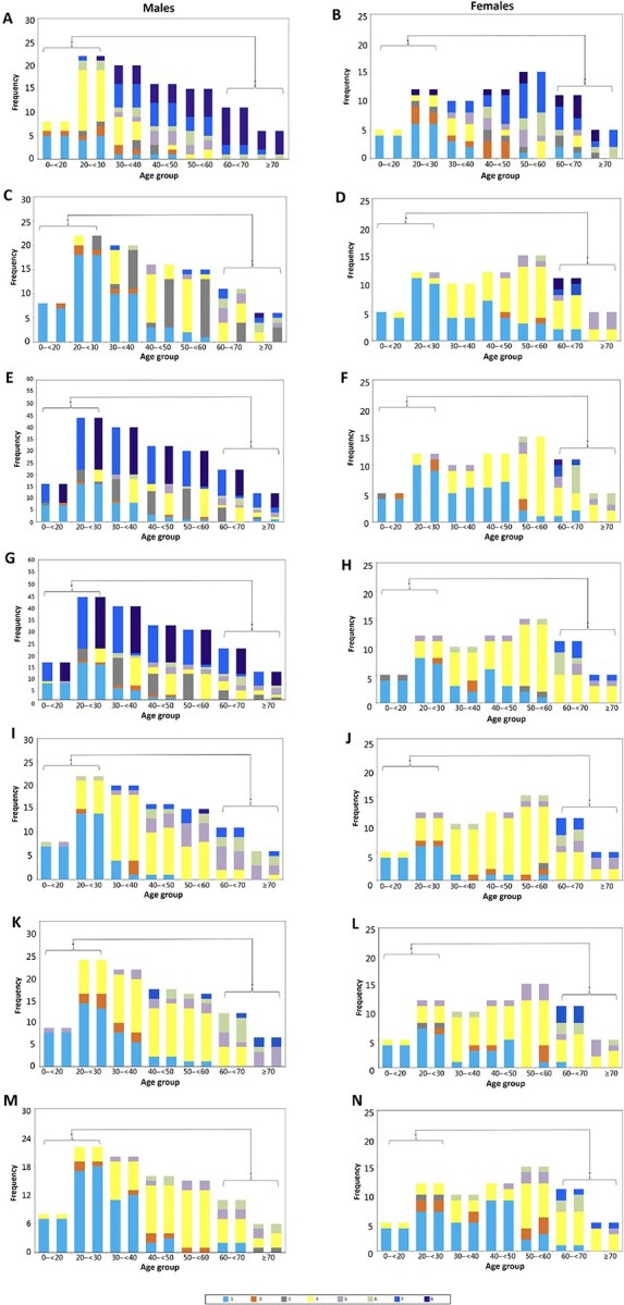
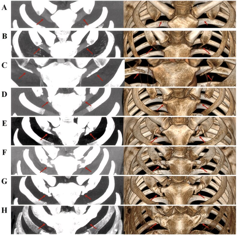
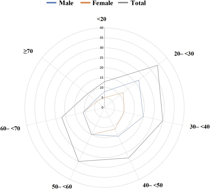
Age estimation of adults is a challenging procedure in forensic practice. Inspired by the previous work by Chinese scholars, we established population-specific age estimation models from the osseous and calcified projections (OCPs) of costal cartilages, using three-dimensional volume-rendering technique. A total of 168 clinical CT scans (2 mm slice thickness) were used to develop the sex-specific age prediction models from a sample of Egyptians, comprising 70 females and 98 males, with documented ages between 12 and 85 years. The sample was also used for validating the Chinese model. We reported the differences between the predictive accuracy of the Egyptian (population specific) and Chinese (non-population specific) models. The most accurate age estimation model was stepwise linear regression with standard error of estimates of 10.9 and 11.8 years in males and females, respectively. For the simple linear regression models, the most accurate formula included OCP of the right second costal cartilage in males and OCP of the left third costal cartilage in females with standard error of estimates of 11.2 and 12.2 years, respectively, and mean absolute error (MAE) of 8.8 and 9.6 years, respectively. By comparison, the best accuracy rates produced by the Chinese vs. the Egyptian models in males and females within 5 years were 30.61% and 32.86% vs. 35.71% and 32.86%, respectively, whereas within 10 years, the accuracy rates increased up to 57.14% and 58.57% vs. 72.45% and 64.29%, respectively. Although the accuracy rates from the Chinese models were lower than those obtained from the Egyptian models, the MAE and least error values were comparable in both sexes. Notable accurate age estimation rates in the advanced age group ≥40 years were reached being 81.25% to 97.92% in males and 69.77% to 93.02% in females. OCP of the right first costal cartilage was the most accurate in cross-population application for males and females with MAE values of 10.7 and 11.03 years, respectively, with balanced accuracy rates of age estimation using the 10-year interval and 40-year cutoff.
Key points: Age differences in calcification form and amount in the seven costal cartilages were found.The best model for males include the second costal cartilage.The best model for females include third or fifth costal cartilages.First OCP is the most accurate in cross-population application regardless of sex.The best OCP in one population is not necessarily the best predictor in both samples.
 求助内容:
求助内容: 应助结果提醒方式:


