{"title":"Neopterin Levels and Immune Response in Autoimmune Uveitis in an Experiment.","authors":"Nadiia Kuryltsiv","doi":"10.3341/kjo.2024.0118","DOIUrl":null,"url":null,"abstract":"<p><strong>Purpose: </strong>To study and compare the immune response and neopterin levels in the blood in experimental autoimmune uveitis (EAU).</p><p><strong>Methods: </strong>A model of EAU was created in 30 Chinchilla rabbits. Intravenous and intravitreal injections of normal horse serum were administered for this purpose. Clinical examinations and blood tests were conducted on days 3, 7, 10, 14, and 21. The blood investigation included the determination of neopterin (NP) level, white blood cell counts, lymphocytes, CD3+, CD4+, CD8+, and CD16+.</p><p><strong>Results: </strong>The peak in white blood cell count was observed on days 7 and 10 (6.4 ± 0.4 g/L and 6.0 ± 0.3 g/L, respectively), lymphocytes on day 3 (68.3% ± 2.4%, 3.0 ± 0.2 g/L), CD3+ on day 7 (64.9% ± 3.1%, 2,032.5 ± 91.2 cells/µL), CD4+ and CD16+ on day 10 (54.6% ± 3.8%, 2,462.3 ± 60.7 cells/µL and 21.8% ± 1.8%, 691.2 ± 37.1 cells/µL, respectively). All these values did not return to the initial ones. There was a gradual decrease in the CD8+ count from day 3 (12.5% ± 1.1%, 142.8 ± 9.1 cells/µL) with a subsequent gradual return towards normal levels by day 21. NP levels incresed on day 3 (5.2 ± 0.7 nmol/L), sustained on day 7 (5.2 ± 0.8 nmol/L), and started to decrease from day 10 (4.25 ± 1.7 nmol/L) to 2.3 ± 0.5 nmol/L on day 21. The highest correlation was observed between clinical manifestations and NP with a correlation coeffient of 0.799 (95% confidence interval, 0.719-0.858), which was significantly stronger (p < 0.05) than the correlations with other immune response markers.</p><p><strong>Conclusions: </strong>During the modeling of EAU, there is an active immune response and a rapid reaction of NP on inflammation. NP is a significantly more sensitive marker of intraocular inflammation than the immune response. It can serve as a predictor of the onset and development of EAU.</p>","PeriodicalId":101356,"journal":{"name":"Korean journal of ophthalmology : KJO","volume":" ","pages":"258-268"},"PeriodicalIF":0.0000,"publicationDate":"2025-06-01","publicationTypes":"Journal Article","fieldsOfStudy":null,"isOpenAccess":false,"openAccessPdf":"https://www.ncbi.nlm.nih.gov/pmc/articles/PMC12178683/pdf/","citationCount":"0","resultStr":null,"platform":"Semanticscholar","paperid":null,"PeriodicalName":"Korean journal of ophthalmology : KJO","FirstCategoryId":"1085","ListUrlMain":"https://doi.org/10.3341/kjo.2024.0118","RegionNum":0,"RegionCategory":null,"ArticlePicture":[],"TitleCN":null,"AbstractTextCN":null,"PMCID":null,"EPubDate":"2025/5/28 0:00:00","PubModel":"Epub","JCR":"","JCRName":"","Score":null,"Total":0}
引用次数: 0
Abstract
Purpose: To study and compare the immune response and neopterin levels in the blood in experimental autoimmune uveitis (EAU).
Methods: A model of EAU was created in 30 Chinchilla rabbits. Intravenous and intravitreal injections of normal horse serum were administered for this purpose. Clinical examinations and blood tests were conducted on days 3, 7, 10, 14, and 21. The blood investigation included the determination of neopterin (NP) level, white blood cell counts, lymphocytes, CD3+, CD4+, CD8+, and CD16+.
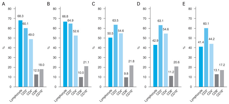
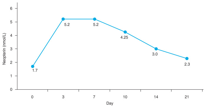
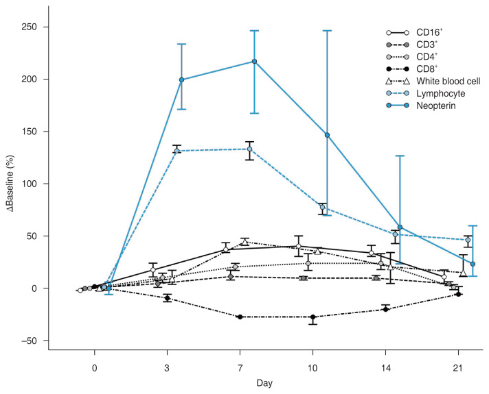
Results: The peak in white blood cell count was observed on days 7 and 10 (6.4 ± 0.4 g/L and 6.0 ± 0.3 g/L, respectively), lymphocytes on day 3 (68.3% ± 2.4%, 3.0 ± 0.2 g/L), CD3+ on day 7 (64.9% ± 3.1%, 2,032.5 ± 91.2 cells/µL), CD4+ and CD16+ on day 10 (54.6% ± 3.8%, 2,462.3 ± 60.7 cells/µL and 21.8% ± 1.8%, 691.2 ± 37.1 cells/µL, respectively). All these values did not return to the initial ones. There was a gradual decrease in the CD8+ count from day 3 (12.5% ± 1.1%, 142.8 ± 9.1 cells/µL) with a subsequent gradual return towards normal levels by day 21. NP levels incresed on day 3 (5.2 ± 0.7 nmol/L), sustained on day 7 (5.2 ± 0.8 nmol/L), and started to decrease from day 10 (4.25 ± 1.7 nmol/L) to 2.3 ± 0.5 nmol/L on day 21. The highest correlation was observed between clinical manifestations and NP with a correlation coeffient of 0.799 (95% confidence interval, 0.719-0.858), which was significantly stronger (p < 0.05) than the correlations with other immune response markers.
Conclusions: During the modeling of EAU, there is an active immune response and a rapid reaction of NP on inflammation. NP is a significantly more sensitive marker of intraocular inflammation than the immune response. It can serve as a predictor of the onset and development of EAU.

 求助内容:
求助内容: 应助结果提醒方式:


