{"title":"Leiomyosarcoma of the Breast: Case Report and Review of the Literature.","authors":"Amal Alimi, Hafedh Abbassi, Skander Abid, Amrou Dinari, Abdeljlil Khlifi, Samir Hidar, Sassi Boughizane, Hedi Khairi","doi":"10.4274/ejbh.galenos.2025.2024-10-8","DOIUrl":null,"url":null,"abstract":"<p><p>Primary breast leiomyosarcoma is an extremely rare malignancy, accounting for approximately 1% of breast tumors and less than 5% of soft tissue sarcomas. Due to its rarity, standardized treatment guidelines remain unclear. We report the case of a 38-year-old woman who presented with a 3 cm, freely mobile breast nodule, initially classified as American College of Radiology Breast Imaging Reporting and Data System 4 on imaging. Core needle biopsy confirmed primary breast leiomyosarcoma, with histopathological and immunohistochemical analysis revealing strong positivity for α-smooth muscle actin, desmin, and H-caldesmon, consistent with smooth muscle differentiation. Epithelial, neural, and vascular markers were negative, ruling out differential diagnoses. The Ki-67 index was 15%, indicating moderate proliferative activity. Staging classified the tumor as T2N0M0 (Stage IIA, the American Joint Committee on Cancer 8<sup>th</sup> edition), and the patient underwent radical mastectomy with sentinel lymph node exploration, followed by adjuvant radiotherapy. Despite the aggressive nature of leiomyosarcomas, this case exhibited favorable prognostic factors, including small tumor size, intermediate grade, negative margins, and no lymphatic spread, suggesting a less aggressive course. After four years of follow-up, the patient remains free of complications, underscoring the importance of long-term monitoring and the need for further research to refine therapeutic approaches.</p>","PeriodicalId":93996,"journal":{"name":"European journal of breast health","volume":" ","pages":"277-280"},"PeriodicalIF":1.7000,"publicationDate":"2025-06-20","publicationTypes":"Journal Article","fieldsOfStudy":null,"isOpenAccess":false,"openAccessPdf":"https://www.ncbi.nlm.nih.gov/pmc/articles/PMC12180097/pdf/","citationCount":"0","resultStr":null,"platform":"Semanticscholar","paperid":null,"PeriodicalName":"European journal of breast health","FirstCategoryId":"1085","ListUrlMain":"https://doi.org/10.4274/ejbh.galenos.2025.2024-10-8","RegionNum":0,"RegionCategory":null,"ArticlePicture":[],"TitleCN":null,"AbstractTextCN":null,"PMCID":null,"EPubDate":"2025/5/29 0:00:00","PubModel":"Epub","JCR":"Q4","JCRName":"ONCOLOGY","Score":null,"Total":0}
引用次数: 0
Abstract
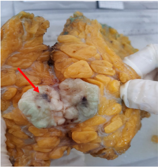
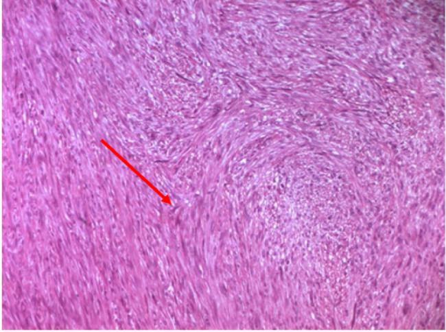
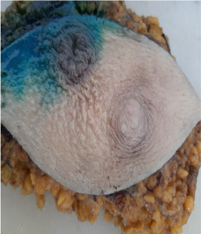
Primary breast leiomyosarcoma is an extremely rare malignancy, accounting for approximately 1% of breast tumors and less than 5% of soft tissue sarcomas. Due to its rarity, standardized treatment guidelines remain unclear. We report the case of a 38-year-old woman who presented with a 3 cm, freely mobile breast nodule, initially classified as American College of Radiology Breast Imaging Reporting and Data System 4 on imaging. Core needle biopsy confirmed primary breast leiomyosarcoma, with histopathological and immunohistochemical analysis revealing strong positivity for α-smooth muscle actin, desmin, and H-caldesmon, consistent with smooth muscle differentiation. Epithelial, neural, and vascular markers were negative, ruling out differential diagnoses. The Ki-67 index was 15%, indicating moderate proliferative activity. Staging classified the tumor as T2N0M0 (Stage IIA, the American Joint Committee on Cancer 8th edition), and the patient underwent radical mastectomy with sentinel lymph node exploration, followed by adjuvant radiotherapy. Despite the aggressive nature of leiomyosarcomas, this case exhibited favorable prognostic factors, including small tumor size, intermediate grade, negative margins, and no lymphatic spread, suggesting a less aggressive course. After four years of follow-up, the patient remains free of complications, underscoring the importance of long-term monitoring and the need for further research to refine therapeutic approaches.

 求助内容:
求助内容: 应助结果提醒方式:


