Luca Albanese, Christoph Germann, Chantal Pauli, Dominic Gascho, Reto Sutter
{"title":"Prevalence and origin of prominent nutrient channels of the ilium bone on MR-imaging.","authors":"Luca Albanese, Christoph Germann, Chantal Pauli, Dominic Gascho, Reto Sutter","doi":"10.1007/s00256-025-04938-x","DOIUrl":null,"url":null,"abstract":"<p><strong>Objective: </strong>Prominent nutrient vessels are commonly seen in the ilium bone, but little is known about their anatomical characteristics. The aim of this study was to investigate the frequency and morphology of these vessels and associated bone-marrow changes of the ilium using MRI.</p><p><strong>Materials and methods: </strong>MRI-examinations of the pelvis in 245 patients were analyzed retrospectively. Prominent nutrient vessels of the ilium were recorded, including vessel origin, anatomical characteristics such as branches, bone-marrow changes, and entry points into the bone.</p><p><strong>Results: </strong>Two hundred forty-five patients (54±16 years, range 18-88, 102 males) were included. Prominent central nutrient vessels were found in virtually all patients on both sides of the ilium. All nutrient vessels arose from the iliolumbar artery, forming a breakthrough-anastomosis to the superior gluteal artery. Two branches were seen in 57.6% on the right and 61.2% on the left side, constituting the most prevalent branching pattern. Three branches were seen in a third for each side. One or four branches were seen in 3-4.5% for both sides. A prominent branching pattern we coined \"central-vessel-convolute\" (CVC) at the central part of the ilium was seen in 75% on either side. Perivascular fatty areas were found in 25% of cases, and in 3.7-2.4% adjacent bone-marrow edema was observed.</p><p><strong>Conclusion: </strong>Prominent nutrient vessels in the ilium are seen in almost all individuals, the majority exhibiting a specific CVC-pattern. These vessels may be surrounded by perivascular fatty areas; adjacent bone-marrow edema is rare. Recognizing the CVCs and the associated imaging findings should facilitate distinguishing normal anatomical structures from pathology.</p>","PeriodicalId":21783,"journal":{"name":"Skeletal Radiology","volume":" ","pages":"2127-2135"},"PeriodicalIF":2.2000,"publicationDate":"2025-10-01","publicationTypes":"Journal Article","fieldsOfStudy":null,"isOpenAccess":false,"openAccessPdf":"https://www.ncbi.nlm.nih.gov/pmc/articles/PMC12361307/pdf/","citationCount":"0","resultStr":null,"platform":"Semanticscholar","paperid":null,"PeriodicalName":"Skeletal Radiology","FirstCategoryId":"3","ListUrlMain":"https://doi.org/10.1007/s00256-025-04938-x","RegionNum":3,"RegionCategory":"医学","ArticlePicture":[],"TitleCN":null,"AbstractTextCN":null,"PMCID":null,"EPubDate":"2025/5/29 0:00:00","PubModel":"Epub","JCR":"Q2","JCRName":"ORTHOPEDICS","Score":null,"Total":0}
引用次数: 0
Abstract
Objective: Prominent nutrient vessels are commonly seen in the ilium bone, but little is known about their anatomical characteristics. The aim of this study was to investigate the frequency and morphology of these vessels and associated bone-marrow changes of the ilium using MRI.
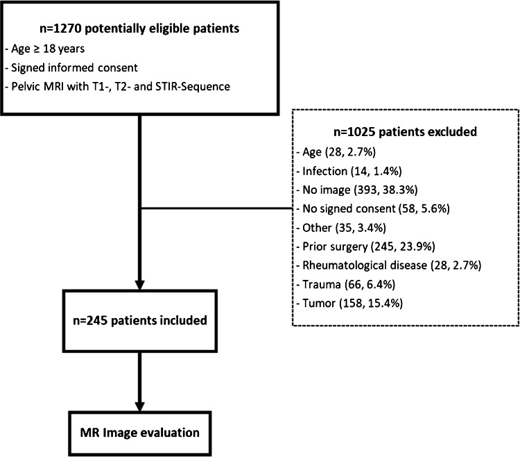
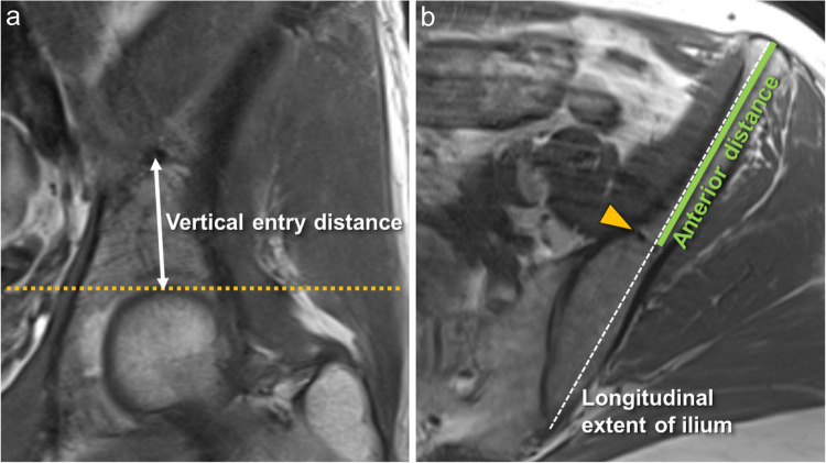
Materials and methods: MRI-examinations of the pelvis in 245 patients were analyzed retrospectively. Prominent nutrient vessels of the ilium were recorded, including vessel origin, anatomical characteristics such as branches, bone-marrow changes, and entry points into the bone.
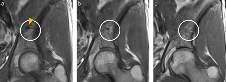
Results: Two hundred forty-five patients (54±16 years, range 18-88, 102 males) were included. Prominent central nutrient vessels were found in virtually all patients on both sides of the ilium. All nutrient vessels arose from the iliolumbar artery, forming a breakthrough-anastomosis to the superior gluteal artery. Two branches were seen in 57.6% on the right and 61.2% on the left side, constituting the most prevalent branching pattern. Three branches were seen in a third for each side. One or four branches were seen in 3-4.5% for both sides. A prominent branching pattern we coined "central-vessel-convolute" (CVC) at the central part of the ilium was seen in 75% on either side. Perivascular fatty areas were found in 25% of cases, and in 3.7-2.4% adjacent bone-marrow edema was observed.
Conclusion: Prominent nutrient vessels in the ilium are seen in almost all individuals, the majority exhibiting a specific CVC-pattern. These vessels may be surrounded by perivascular fatty areas; adjacent bone-marrow edema is rare. Recognizing the CVCs and the associated imaging findings should facilitate distinguishing normal anatomical structures from pathology.
期刊介绍:
Skeletal Radiology provides a forum for the dissemination of current knowledge and information dealing with disorders of the musculoskeletal system including the spine. While emphasizing the radiological aspects of the many varied skeletal abnormalities, the journal also adopts an interdisciplinary approach, reflecting the membership of the International Skeletal Society. Thus, the anatomical, pathological, physiological, clinical, metabolic and epidemiological aspects of the many entities affecting the skeleton receive appropriate consideration.
This is the Journal of the International Skeletal Society and the Official Journal of the Society of Skeletal Radiology and the Australasian Musculoskelelal Imaging Group.

 求助内容:
求助内容: 应助结果提醒方式:


