[68Ga]Ga-PSMA-11 PET/CT and [18F]Fluorocholine PET/CT in Assessment and Clinical Decision Making of Recurrent Prostate Cancer: A Prospective Crossover Trial.
IF 2.5 4区 医学Q2 RADIOLOGY, NUCLEAR MEDICINE & MEDICAL IMAGING
Mohsen Beheshti, Malihe Shahbazi-Akbari, Marcus Hacker, Wolfgang Loidl, Werner Langsteger
{"title":"[<sup>68</sup>Ga]Ga-PSMA-11 PET/CT and [<sup>18</sup>F]Fluorocholine PET/CT in Assessment and Clinical Decision Making of Recurrent Prostate Cancer: A Prospective Crossover Trial.","authors":"Mohsen Beheshti, Malihe Shahbazi-Akbari, Marcus Hacker, Wolfgang Loidl, Werner Langsteger","doi":"10.1007/s11307-025-02020-5","DOIUrl":null,"url":null,"abstract":"<p><strong>Purpose: </strong>There are few prospective studies addressed toward the role of <sup>68</sup>Gallium-labelled prostate-specific membrane antigen-11 ([<sup>68</sup>Ga]Ga-PSMA-11) compared to [<sup>18</sup>F]Fluorocholine ([<sup>18</sup>F]FCH) PET/CT in clinical decision-making as prostate-specific PET-tracers. This study aims to evaluate the impact of PET/CT using [<sup>68</sup>Ga]Ga-PSMA-11 and [<sup>18</sup>F]FCH in clinical management of recurrent prostate cancer (PCa) and correlates imaging findings with clinical characteristics of PCa.</p><p><strong>Procedures: </strong>Forty-six patients with PCa (mean age 68.3 ± 6.3 years) with biochemical recurrence were enrolled in this prospective crossover trial. All patients underwent both [<sup>68</sup>Ga]Ga-PSMA-11 and [<sup>18</sup>F]FCH PET/CT within a maximum interval of 12 days (median 7d). A standard randomization tool randomized the sequence of PET/CT imaging. Clinical decision-making occurred in an interdisciplinary meeting considering PET/CT findings. PET/CT-blinded readings were performed 3 months after imaging followed by a consensus meeting for final interpretation of detected lesions.</p><p><strong>Results: </strong>Both imaging modalities detected 136 total malignant lesions. [<sup>68</sup>Ga]Ga-PSMA-11 and [<sup>18</sup>F]FCH PET/CT detected 125 and 60 lesions with a sensitivity of 96% and 48%, respectively. Tumor-to-background ratios and semi-quantitative PET parameters on [<sup>68</sup>Ga]Ga-PSMA-11 were significantly higher in 54 (41.2%) tracer-avid congruent lesions detected on both imaging modalities. [<sup>68</sup>Ga]Ga-PSMA-11 PET/CT exclusively detected 71 (52.2%) lesions, while 6 (4.4%) lesions were solely seen on [<sup>18</sup>F]FCH PET/CT. [<sup>68</sup>Ga]Ga-PSMA-11 and [<sup>18</sup>F]FCH PET/CT were positive in 35/46 (76%) and 26/46 (57%) patients, respectively. PET/CT imaging led to a major treatment change in 4 (8.7%) patients, of which [<sup>18</sup>F]FCH PET/CT had superior impact in one patient.</p><p><strong>Conclusions: </strong>[<sup>68</sup>Ga]Ga-PSMA-11 PET/CT revealed superior diagnostic performance to [<sup>18</sup>F]FCH PET/CT in patients with recurrent PCa, specifically with very low PSA levels ≤ 1 ng/ml. Moreover, it led to more accurate staging and clinical management of the disease. [<sup>18</sup>F]FCH PET/CT may play a complementary role in rare, select high-risk cases with negative [<sup>68</sup>Ga]Ga-PSMA-11 PET/CT and ongoing ADT.</p>","PeriodicalId":18760,"journal":{"name":"Molecular Imaging and Biology","volume":" ","pages":"597-605"},"PeriodicalIF":2.5000,"publicationDate":"2025-08-01","publicationTypes":"Journal Article","fieldsOfStudy":null,"isOpenAccess":false,"openAccessPdf":"https://www.ncbi.nlm.nih.gov/pmc/articles/PMC12405339/pdf/","citationCount":"0","resultStr":null,"platform":"Semanticscholar","paperid":null,"PeriodicalName":"Molecular Imaging and Biology","FirstCategoryId":"3","ListUrlMain":"https://doi.org/10.1007/s11307-025-02020-5","RegionNum":4,"RegionCategory":"医学","ArticlePicture":[],"TitleCN":null,"AbstractTextCN":null,"PMCID":null,"EPubDate":"2025/5/28 0:00:00","PubModel":"Epub","JCR":"Q2","JCRName":"RADIOLOGY, NUCLEAR MEDICINE & MEDICAL IMAGING","Score":null,"Total":0}
引用次数: 0
Abstract
Purpose: There are few prospective studies addressed toward the role of 68Gallium-labelled prostate-specific membrane antigen-11 ([68Ga]Ga-PSMA-11) compared to [18F]Fluorocholine ([18F]FCH) PET/CT in clinical decision-making as prostate-specific PET-tracers. This study aims to evaluate the impact of PET/CT using [68Ga]Ga-PSMA-11 and [18F]FCH in clinical management of recurrent prostate cancer (PCa) and correlates imaging findings with clinical characteristics of PCa.
Procedures: Forty-six patients with PCa (mean age 68.3 ± 6.3 years) with biochemical recurrence were enrolled in this prospective crossover trial. All patients underwent both [68Ga]Ga-PSMA-11 and [18F]FCH PET/CT within a maximum interval of 12 days (median 7d). A standard randomization tool randomized the sequence of PET/CT imaging. Clinical decision-making occurred in an interdisciplinary meeting considering PET/CT findings. PET/CT-blinded readings were performed 3 months after imaging followed by a consensus meeting for final interpretation of detected lesions.
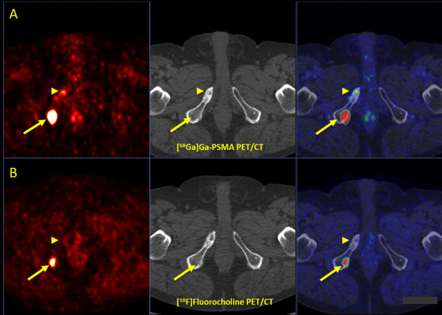
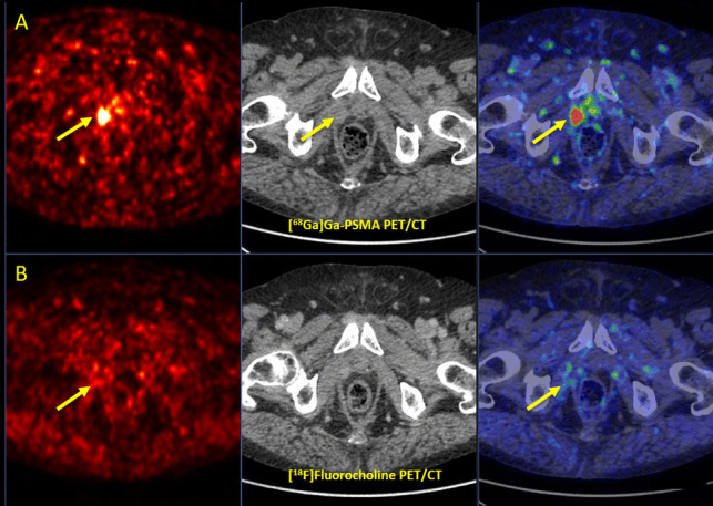
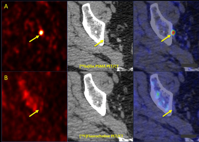
Results: Both imaging modalities detected 136 total malignant lesions. [68Ga]Ga-PSMA-11 and [18F]FCH PET/CT detected 125 and 60 lesions with a sensitivity of 96% and 48%, respectively. Tumor-to-background ratios and semi-quantitative PET parameters on [68Ga]Ga-PSMA-11 were significantly higher in 54 (41.2%) tracer-avid congruent lesions detected on both imaging modalities. [68Ga]Ga-PSMA-11 PET/CT exclusively detected 71 (52.2%) lesions, while 6 (4.4%) lesions were solely seen on [18F]FCH PET/CT. [68Ga]Ga-PSMA-11 and [18F]FCH PET/CT were positive in 35/46 (76%) and 26/46 (57%) patients, respectively. PET/CT imaging led to a major treatment change in 4 (8.7%) patients, of which [18F]FCH PET/CT had superior impact in one patient.
Conclusions: [68Ga]Ga-PSMA-11 PET/CT revealed superior diagnostic performance to [18F]FCH PET/CT in patients with recurrent PCa, specifically with very low PSA levels ≤ 1 ng/ml. Moreover, it led to more accurate staging and clinical management of the disease. [18F]FCH PET/CT may play a complementary role in rare, select high-risk cases with negative [68Ga]Ga-PSMA-11 PET/CT and ongoing ADT.
期刊介绍:
Molecular Imaging and Biology (MIB) invites original contributions (research articles, review articles, commentaries, etc.) on the utilization of molecular imaging (i.e., nuclear imaging, optical imaging, autoradiography and pathology, MRI, MPI, ultrasound imaging, radiomics/genomics etc.) to investigate questions related to biology and health. The objective of MIB is to provide a forum to the discovery of molecular mechanisms of disease through the use of imaging techniques. We aim to investigate the biological nature of disease in patients and establish new molecular imaging diagnostic and therapy procedures.
Some areas that are covered are:
Preclinical and clinical imaging of macromolecular targets (e.g., genes, receptors, enzymes) involved in significant biological processes.
The design, characterization, and study of new molecular imaging probes and contrast agents for the functional interrogation of macromolecular targets.
Development and evaluation of imaging systems including instrumentation, image reconstruction algorithms, image analysis, and display.
Development of molecular assay approaches leading to quantification of the biological information obtained in molecular imaging.
Study of in vivo animal models of disease for the development of new molecular diagnostics and therapeutics.
Extension of in vitro and in vivo discoveries using disease models, into well designed clinical research investigations.
Clinical molecular imaging involving clinical investigations, clinical trials and medical management or cost-effectiveness studies.

 求助内容:
求助内容: 应助结果提醒方式:


