Roberto Manfrellotti, Dario Gagliano, Roberta Costanzo, Alejandra Mosteiro, Marta Codes Méndez, Doriam Perera Valdivia, Nikolay Lasunin, Carlo Giorgio Giussani, Giorgio Giovanni Carrabba, Joaquim Enseñat, Alberto Di Somma, Alberto Prats-Galino
{"title":"The meningo-orbital band from an endoscopic transorbital approach: an anatomical study.","authors":"Roberto Manfrellotti, Dario Gagliano, Roberta Costanzo, Alejandra Mosteiro, Marta Codes Méndez, Doriam Perera Valdivia, Nikolay Lasunin, Carlo Giorgio Giussani, Giorgio Giovanni Carrabba, Joaquim Enseñat, Alberto Di Somma, Alberto Prats-Galino","doi":"10.3389/fnana.2025.1578959","DOIUrl":null,"url":null,"abstract":"<p><strong>Introduction: </strong>The meningo-orbital band (MOB) is an intricate dural structure extending between the periorbita, the frontal dura, and the temporal dura. The endoscopic transorbital approach (ETOA) provides a more thorough understanding of its anatomy.</p><p><strong>Materials and methods: </strong>Anatomical dissections were performed on 15 human head specimens (30 orbits) at the Laboratory of Surgical Neuroanatomy (LSNA) at the University of Barcelona. The specimens were preserved using a Cambridge solution for optimal fixation. An endoscopic transorbital approach (ETOA) was used to isolate the meningo-orbital band (MOB). A rigid 4-mm endoscope with an HD camera and light source was used for the procedure. Multislice helical CT scans were performed both before and after the dissections to document the anatomical features. Additionally, a specialized software (The ImagingSource®) was used to calculate the variability in the angle between the first two bone pillars of the ETOA: the sagittal crest (SC) and the lesser sphenoid wing (LSW). The vascularization of the MOB was studied by longitudinally cutting the band and using red and blue latex injections into the carotid arteries and jugular veins, respectively, to highlight the cerebral vasculature.</p><p><strong>Results: </strong>In the endoscopic transorbital approach (ETOA), key structures, including the greater and lesser sphenoid wings, are excised, exposing the meningo-orbital band (MOB). The MOB extends from the periorbita medially to the frontal and temporal dura laterally and is firmly attached to the anterior clinoid process (ACP). Anatomical dissection reveals the MOB's complex three-dimensional structure and its relationships with cranial nerves III, IV, and V1 along the lateral wall of the cavernous sinus and the superior orbital fissure (SOF). The ACP serves as a protective barrier between the MOB and the paraclinoid segment of the internal carotid artery (ICA). Additionally, the MOB is vascularized by the MOB artery (MOBA), a branch of the middle meningeal artery, which bifurcates into the frontal and temporal branches.</p><p><strong>Conclusion: </strong>This study highlights the key anatomical relationships of the meningo-orbital band (MOB) with critical structures, including cranial nerves III, IV, and V1, as well as the ICA. These findings are essential for refining surgical planning and improving the safety and precision of skull base surgery.</p>","PeriodicalId":12572,"journal":{"name":"Frontiers in Neuroanatomy","volume":"19 ","pages":"1578959"},"PeriodicalIF":2.3000,"publicationDate":"2025-05-14","publicationTypes":"Journal Article","fieldsOfStudy":null,"isOpenAccess":false,"openAccessPdf":"https://www.ncbi.nlm.nih.gov/pmc/articles/PMC12116505/pdf/","citationCount":"0","resultStr":null,"platform":"Semanticscholar","paperid":null,"PeriodicalName":"Frontiers in Neuroanatomy","FirstCategoryId":"3","ListUrlMain":"https://doi.org/10.3389/fnana.2025.1578959","RegionNum":4,"RegionCategory":"医学","ArticlePicture":[],"TitleCN":null,"AbstractTextCN":null,"PMCID":null,"EPubDate":"2025/1/1 0:00:00","PubModel":"eCollection","JCR":"Q1","JCRName":"ANATOMY & MORPHOLOGY","Score":null,"Total":0}
引用次数: 0
Abstract
Introduction: The meningo-orbital band (MOB) is an intricate dural structure extending between the periorbita, the frontal dura, and the temporal dura. The endoscopic transorbital approach (ETOA) provides a more thorough understanding of its anatomy.
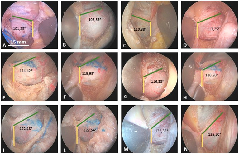
Materials and methods: Anatomical dissections were performed on 15 human head specimens (30 orbits) at the Laboratory of Surgical Neuroanatomy (LSNA) at the University of Barcelona. The specimens were preserved using a Cambridge solution for optimal fixation. An endoscopic transorbital approach (ETOA) was used to isolate the meningo-orbital band (MOB). A rigid 4-mm endoscope with an HD camera and light source was used for the procedure. Multislice helical CT scans were performed both before and after the dissections to document the anatomical features. Additionally, a specialized software (The ImagingSource®) was used to calculate the variability in the angle between the first two bone pillars of the ETOA: the sagittal crest (SC) and the lesser sphenoid wing (LSW). The vascularization of the MOB was studied by longitudinally cutting the band and using red and blue latex injections into the carotid arteries and jugular veins, respectively, to highlight the cerebral vasculature.
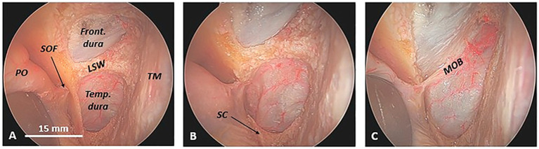
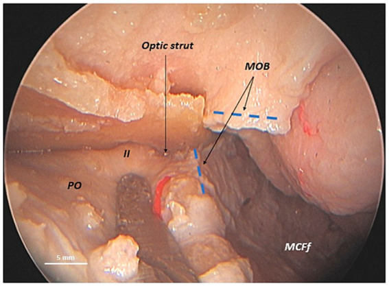
Results: In the endoscopic transorbital approach (ETOA), key structures, including the greater and lesser sphenoid wings, are excised, exposing the meningo-orbital band (MOB). The MOB extends from the periorbita medially to the frontal and temporal dura laterally and is firmly attached to the anterior clinoid process (ACP). Anatomical dissection reveals the MOB's complex three-dimensional structure and its relationships with cranial nerves III, IV, and V1 along the lateral wall of the cavernous sinus and the superior orbital fissure (SOF). The ACP serves as a protective barrier between the MOB and the paraclinoid segment of the internal carotid artery (ICA). Additionally, the MOB is vascularized by the MOB artery (MOBA), a branch of the middle meningeal artery, which bifurcates into the frontal and temporal branches.
Conclusion: This study highlights the key anatomical relationships of the meningo-orbital band (MOB) with critical structures, including cranial nerves III, IV, and V1, as well as the ICA. These findings are essential for refining surgical planning and improving the safety and precision of skull base surgery.
期刊介绍:
Frontiers in Neuroanatomy publishes rigorously peer-reviewed research revealing important aspects of the anatomical organization of all nervous systems across all species. Specialty Chief Editor Javier DeFelipe at the Cajal Institute (CSIC) is supported by an outstanding Editorial Board of international experts. This multidisciplinary open-access journal is at the forefront of disseminating and communicating scientific knowledge and impactful discoveries to researchers, academics, clinicians and the public worldwide.
 求助内容:
求助内容: 应助结果提醒方式:


