Xinyi Chen, Yanfeng Zhao, Yunchong Han, Kai Wei, Shufang Cheng, Yongjun Ye, Jie Feng, Xinchen Huang, Jingjing Xu
{"title":"A diagnostic model based on magnetic resonance imaging for Menière’s disease: a multicentre study","authors":"Xinyi Chen, Yanfeng Zhao, Yunchong Han, Kai Wei, Shufang Cheng, Yongjun Ye, Jie Feng, Xinchen Huang, Jingjing Xu","doi":"10.4274/dir.2025.253293","DOIUrl":null,"url":null,"abstract":"<p><strong>Purpose: </strong>To evaluate the diagnostic performance of delayed post-gadolinium enhancement magnetic resonance imaging (DEMRI) in diagnosing Menière’s disease (MD) and to establish an effective MRI-based diagnostic model.</p><p><strong>Methods: </strong>This retrospective multicenter study assessed DEMRI descriptors in patients presenting with Ménièriform symptoms who were examined consecutively between May 2022 and May 2024. A total of 162 ears (95 with MD, 67 controls) were included. Each ear was randomly assigned to either a training set (n = 98) or a validation set (n = 64). In the training cohort, diagnostic models for MD were developed using logistic regression. The area under the curve (AUC) was used to evaluate the diagnostic performance of the different models. The Delong test was applied to compare AUC estimates between models.</p><p><strong>Results: </strong>The proposed DEMRI diagnostic model demonstrated strong diagnostic performance in both the training cohort (AUC: 0.907) and the validation cohort (AUC: 0.887), outperforming the clinical diagnostic model (<i>P</i> = 0.01231; 95% confidence interval: 0.033–0.269) in the validation cohort. The AUC of the DEMRI model was also higher than that of the combined DEMRI-clinical model (AUC: 0.796), although the difference was not statistically significant (<i>P</i> = 0.054). In the training set, the sensitivity and specificity of the DEMRI model were 78.9% and 88.5%, respectively.</p><p><strong>Conclusion: </strong>A diagnostic model based on DEMRI features for MD is more effective than one based solely on clinical variables. DEMRI should, therefore, be recommended when MD is suspected, given its significant diagnostic potential.</p><p><strong>Clinical significance: </strong>This model may improve the accuracy and timeliness of MD diagnosis, as it is less influenced by the attending physician’s level of inquiry or the patient’s self-reporting ability. It may also contribute to more effective disease management in patients with MD.</p>","PeriodicalId":11341,"journal":{"name":"Diagnostic and interventional radiology","volume":" ","pages":"347-358"},"PeriodicalIF":1.7000,"publicationDate":"2025-07-08","publicationTypes":"Journal Article","fieldsOfStudy":null,"isOpenAccess":false,"openAccessPdf":"https://www.ncbi.nlm.nih.gov/pmc/articles/PMC12239533/pdf/","citationCount":"0","resultStr":null,"platform":"Semanticscholar","paperid":null,"PeriodicalName":"Diagnostic and interventional radiology","FirstCategoryId":"3","ListUrlMain":"https://doi.org/10.4274/dir.2025.253293","RegionNum":4,"RegionCategory":"医学","ArticlePicture":[],"TitleCN":null,"AbstractTextCN":null,"PMCID":null,"EPubDate":"2025/5/29 0:00:00","PubModel":"Epub","JCR":"Q3","JCRName":"RADIOLOGY, NUCLEAR MEDICINE & MEDICAL IMAGING","Score":null,"Total":0}
引用次数: 0
Abstract
Purpose: To evaluate the diagnostic performance of delayed post-gadolinium enhancement magnetic resonance imaging (DEMRI) in diagnosing Menière’s disease (MD) and to establish an effective MRI-based diagnostic model.
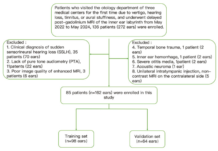
Methods: This retrospective multicenter study assessed DEMRI descriptors in patients presenting with Ménièriform symptoms who were examined consecutively between May 2022 and May 2024. A total of 162 ears (95 with MD, 67 controls) were included. Each ear was randomly assigned to either a training set (n = 98) or a validation set (n = 64). In the training cohort, diagnostic models for MD were developed using logistic regression. The area under the curve (AUC) was used to evaluate the diagnostic performance of the different models. The Delong test was applied to compare AUC estimates between models.
Results: The proposed DEMRI diagnostic model demonstrated strong diagnostic performance in both the training cohort (AUC: 0.907) and the validation cohort (AUC: 0.887), outperforming the clinical diagnostic model (P = 0.01231; 95% confidence interval: 0.033–0.269) in the validation cohort. The AUC of the DEMRI model was also higher than that of the combined DEMRI-clinical model (AUC: 0.796), although the difference was not statistically significant (P = 0.054). In the training set, the sensitivity and specificity of the DEMRI model were 78.9% and 88.5%, respectively.
Conclusion: A diagnostic model based on DEMRI features for MD is more effective than one based solely on clinical variables. DEMRI should, therefore, be recommended when MD is suspected, given its significant diagnostic potential.
Clinical significance: This model may improve the accuracy and timeliness of MD diagnosis, as it is less influenced by the attending physician’s level of inquiry or the patient’s self-reporting ability. It may also contribute to more effective disease management in patients with MD.
期刊介绍:
Diagnostic and Interventional Radiology (Diagn Interv Radiol) is the open access, online-only official publication of Turkish Society of Radiology. It is published bimonthly and the journal’s publication language is English.
The journal is a medium for original articles, reviews, pictorial essays, technical notes related to all fields of diagnostic and interventional radiology.

 求助内容:
求助内容: 应助结果提醒方式:


