{"title":"Cerebral blood flow based on 3D-ASL technology in the early detection of mild cognitive impairment in type 2 diabetic patients: a pilot study.","authors":"Tingzhou Hou, Yijie Wang, Hanwen Xu, Zhenyu Shu, Xiaodong He, Xiaohong Wu","doi":"10.3389/fendo.2025.1576998","DOIUrl":null,"url":null,"abstract":"<p><strong>Context: </strong>Type 2 diabetes mellitus (T2DM) significantly increases the risk of mild cognitive impairment (MCI), and earlier recognition is crucial for timely intervention and improving patients' quality of life.</p><p><strong>Objective: </strong>The aim of this study was to characterize changes in cerebral white matter hyperintensities (WMH) and cerebral blood flow (CBF) based on 3D-arterial spin labeling (3D-ASL) measurements in T2DM with MCI, and to assess their potential as markers for early prediction of MCI.</p><p><strong>Methods: </strong>This study included 30 T2DM patients stratified into T2DM-MCI and T2DM-nMCI groups using MMSE/MoCA. MRI assessed cerebral WMH volume (T2-FLAIR) and regional CBF (3D-ASL) in temporal, parietal, occipital, and hippocampal areas. Group differences in WMH/CBF were analyzed, ROC evaluated CBF's diagnostic efficacy for MCI, and CBF-cognition correlations were assessed via Spearman's analysis.</p><p><strong>Results: </strong>Cranial MRI analysis shows that there were no significant differences between the two groups in terms of total cerebral WMH volume and regional volume (P>0.05). CBF in the temporal, parietal, occipital, and hippocampal regions was significantly lower in the T2DM-MCI group than in the T2DM-nMCI group (P<0.05). ROC analysis revealed that CBF in the hippocampus had the highest diagnostic efficacy (AUC = 0.813, optimal cutoff value of 41.165 ml/(100 g·min), sensitivity 73.3%, specificity 80.0%). Spearman's correlation analysis showed that CBF in the temporal, parietal, occipital, and hippocampal regions was significantly and positively correlated with MoCA scores (P < 0.05), with temporal and parietal CBF also significantly and positively correlated with MMSE scores (P < 0.05).</p><p><strong>Conclusions: </strong>CBF, based on 3D-ASL measurements, especially in the hippocampus, is a promising functional marker for identifying MCI in patients with T2DM.</p>","PeriodicalId":12447,"journal":{"name":"Frontiers in Endocrinology","volume":"16 ","pages":"1576998"},"PeriodicalIF":4.6000,"publicationDate":"2025-05-14","publicationTypes":"Journal Article","fieldsOfStudy":null,"isOpenAccess":false,"openAccessPdf":"https://www.ncbi.nlm.nih.gov/pmc/articles/PMC12116335/pdf/","citationCount":"0","resultStr":null,"platform":"Semanticscholar","paperid":null,"PeriodicalName":"Frontiers in Endocrinology","FirstCategoryId":"3","ListUrlMain":"https://doi.org/10.3389/fendo.2025.1576998","RegionNum":2,"RegionCategory":"医学","ArticlePicture":[],"TitleCN":null,"AbstractTextCN":null,"PMCID":null,"EPubDate":"2025/1/1 0:00:00","PubModel":"eCollection","JCR":"Q2","JCRName":"ENDOCRINOLOGY & METABOLISM","Score":null,"Total":0}
引用次数: 0
Abstract
Context: Type 2 diabetes mellitus (T2DM) significantly increases the risk of mild cognitive impairment (MCI), and earlier recognition is crucial for timely intervention and improving patients' quality of life.
Objective: The aim of this study was to characterize changes in cerebral white matter hyperintensities (WMH) and cerebral blood flow (CBF) based on 3D-arterial spin labeling (3D-ASL) measurements in T2DM with MCI, and to assess their potential as markers for early prediction of MCI.
Methods: This study included 30 T2DM patients stratified into T2DM-MCI and T2DM-nMCI groups using MMSE/MoCA. MRI assessed cerebral WMH volume (T2-FLAIR) and regional CBF (3D-ASL) in temporal, parietal, occipital, and hippocampal areas. Group differences in WMH/CBF were analyzed, ROC evaluated CBF's diagnostic efficacy for MCI, and CBF-cognition correlations were assessed via Spearman's analysis.
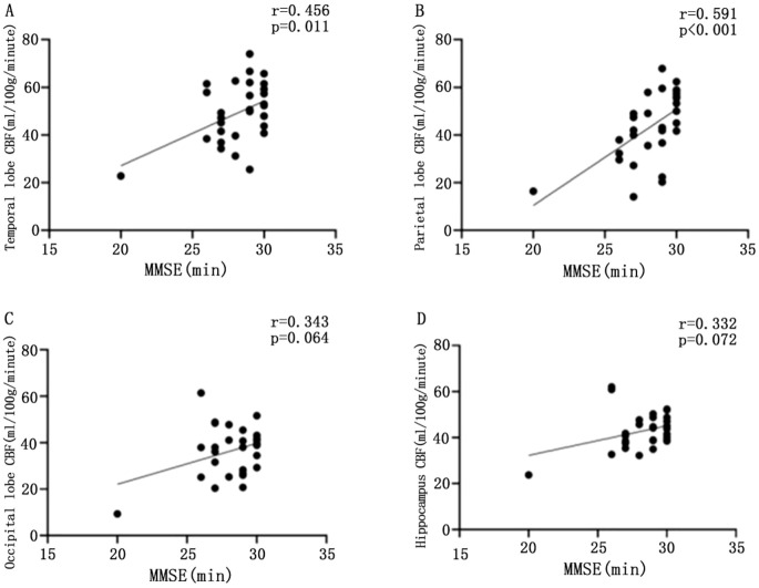
Results: Cranial MRI analysis shows that there were no significant differences between the two groups in terms of total cerebral WMH volume and regional volume (P>0.05). CBF in the temporal, parietal, occipital, and hippocampal regions was significantly lower in the T2DM-MCI group than in the T2DM-nMCI group (P<0.05). ROC analysis revealed that CBF in the hippocampus had the highest diagnostic efficacy (AUC = 0.813, optimal cutoff value of 41.165 ml/(100 g·min), sensitivity 73.3%, specificity 80.0%). Spearman's correlation analysis showed that CBF in the temporal, parietal, occipital, and hippocampal regions was significantly and positively correlated with MoCA scores (P < 0.05), with temporal and parietal CBF also significantly and positively correlated with MMSE scores (P < 0.05).
Conclusions: CBF, based on 3D-ASL measurements, especially in the hippocampus, is a promising functional marker for identifying MCI in patients with T2DM.
期刊介绍:
Frontiers in Endocrinology is a field journal of the "Frontiers in" journal series.
In today’s world, endocrinology is becoming increasingly important as it underlies many of the challenges societies face - from obesity and diabetes to reproduction, population control and aging. Endocrinology covers a broad field from basic molecular and cellular communication through to clinical care and some of the most crucial public health issues. The journal, thus, welcomes outstanding contributions in any domain of endocrinology.
Frontiers in Endocrinology publishes articles on the most outstanding discoveries across a wide research spectrum of Endocrinology. The mission of Frontiers in Endocrinology is to bring all relevant Endocrinology areas together on a single platform.


 求助内容:
求助内容: 应助结果提醒方式:


