Débora Costa Ruiz, Rocharles Cavalcante Fontenele, Amanda Farias-Gomes, Hugo Gaêta-Araujo, Matheus Lima Oliveira, Deborah Queiroz Freitas, Francisco Haiter-Neto
{"title":"Influence of a portable X-ray device in the diagnosis of proximal caries lesions.","authors":"Débora Costa Ruiz, Rocharles Cavalcante Fontenele, Amanda Farias-Gomes, Hugo Gaêta-Araujo, Matheus Lima Oliveira, Deborah Queiroz Freitas, Francisco Haiter-Neto","doi":"10.1590/1807-3107bor-2025.vol39.028","DOIUrl":null,"url":null,"abstract":"<p><p>This study aimed to evaluate the influence of a portable X-ray device on the diagnosis of proximal caries lesions. For that, radiographs of 40 human teeth with white spots or color changes in enamel and/or dentin were acquired using the Eagle X-ray portable device (Alliage, São Paulo, Brazil) set at 2.5 mA, 60 kVp and an exposure time of 0.5 s (1.25 mAs). Then, new radiographs of the teeth were acquired using the Focus X-ray wall-mounted device (Instrumentarium, Tuusula, Finland) set at 7 mA, 70 kVp, and exposure time of 0.16 s (1.12 mAs). Five oral and maxillofacial radiologists individually assessed the radiographs. Area under the receiver operating characteristic curve (AUC), sensitivity, and specificity were calculated from the responses of the five examiners and compared between the devices tested using Student's t test. Significance level was set at 5% (α = 0.05). The weighted Kappa index evaluated the intra- and inter-examiner agreements for caries lesions diagnosis. The use of a portable X-ray device did not influence on AUC, sensitivity and specificity metrics for the diagnosis of caries lesions (p > 0.05). The intra- and inter-examiner agreements for the caries lesions diagnosis ranged from substantial to almost perfect (0.646-0.859) and moderate to substantial (0.491-0.617), respectively. The diagnostic accuracy for detecting proximal caries lesions is not impaired when using a portable X-ray device.</p>","PeriodicalId":9240,"journal":{"name":"Brazilian oral research","volume":"39 ","pages":"e028"},"PeriodicalIF":1.3000,"publicationDate":"2025-05-23","publicationTypes":"Journal Article","fieldsOfStudy":null,"isOpenAccess":false,"openAccessPdf":"https://www.ncbi.nlm.nih.gov/pmc/articles/PMC12108113/pdf/","citationCount":"0","resultStr":null,"platform":"Semanticscholar","paperid":null,"PeriodicalName":"Brazilian oral research","FirstCategoryId":"3","ListUrlMain":"https://doi.org/10.1590/1807-3107bor-2025.vol39.028","RegionNum":4,"RegionCategory":"医学","ArticlePicture":[],"TitleCN":null,"AbstractTextCN":null,"PMCID":null,"EPubDate":"2025/1/1 0:00:00","PubModel":"eCollection","JCR":"Q3","JCRName":"DENTISTRY, ORAL SURGERY & MEDICINE","Score":null,"Total":0}
引用次数: 0
Abstract
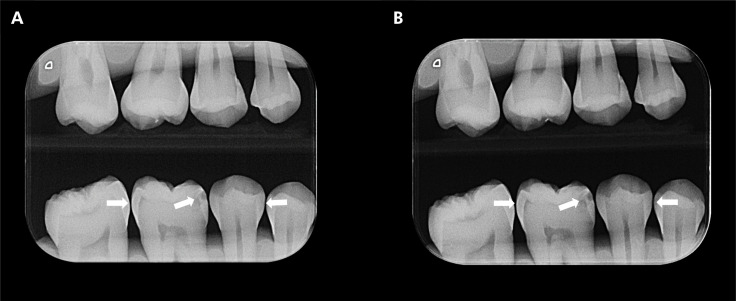
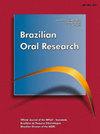
This study aimed to evaluate the influence of a portable X-ray device on the diagnosis of proximal caries lesions. For that, radiographs of 40 human teeth with white spots or color changes in enamel and/or dentin were acquired using the Eagle X-ray portable device (Alliage, São Paulo, Brazil) set at 2.5 mA, 60 kVp and an exposure time of 0.5 s (1.25 mAs). Then, new radiographs of the teeth were acquired using the Focus X-ray wall-mounted device (Instrumentarium, Tuusula, Finland) set at 7 mA, 70 kVp, and exposure time of 0.16 s (1.12 mAs). Five oral and maxillofacial radiologists individually assessed the radiographs. Area under the receiver operating characteristic curve (AUC), sensitivity, and specificity were calculated from the responses of the five examiners and compared between the devices tested using Student's t test. Significance level was set at 5% (α = 0.05). The weighted Kappa index evaluated the intra- and inter-examiner agreements for caries lesions diagnosis. The use of a portable X-ray device did not influence on AUC, sensitivity and specificity metrics for the diagnosis of caries lesions (p > 0.05). The intra- and inter-examiner agreements for the caries lesions diagnosis ranged from substantial to almost perfect (0.646-0.859) and moderate to substantial (0.491-0.617), respectively. The diagnostic accuracy for detecting proximal caries lesions is not impaired when using a portable X-ray device.
 求助内容:
求助内容: 应助结果提醒方式:


