{"title":"Optical coherence tomography retinal imaging: narrative review of technological advancements and clinical applications.","authors":"Christopher S Langlo, Aana Amin, Susanna S Park","doi":"10.21037/atm-24-211","DOIUrl":null,"url":null,"abstract":"<p><strong>Background and objective: </strong>Optical coherence tomography (OCT) is a non-invasive imaging tool that can provide rapid cross-sectional images of the retina, cornea, and optic nerve head in live patients. The objective of this review is to provide an overview of the technical advancements and current clinical applications of OCT for managing patients with retinal disorders.</p><p><strong>Methods: </strong>Narrative overview synthesizing the findings of literature retrieved from searches of computerized database, authoritative texts and authors' clinical experience and expertise.</p><p><strong>Key content and findings: </strong>Unlike the first-generation time-domain OCT (TD-OCT) instruments, the newer spectral-domain OCT (SD-OCT) instruments use a broadband light source to increase axial image resolution. In addition, the decreased image acquisition time also increases the transverse image resolution, reduces motion artifacts, and allows serial cross-sectional images of the retina to be obtained rapidly. A three-dimensional (3D) image of the retina, reconstructed using serial two-dimensional (2D) OCT images, can be used to quantitate retinal thickness and volume and perform analysis of retinal topography. Currently, commercial SD-OCT instruments are used routinely in clinical practice to obtain morphologic information used to diagnose and manage patients with various retinal disorders including macular degeneration and diabetic retinopathy. Newer swept-source OCT technology with faster image acquisition, provides wider field imaging of the peripheral retina. SD-OCT instruments can be incorporated into surgical microscopes to allow imaging of the retina during retinal surgery so that morphologic changes in the retina from surgical maneuvers can be observed in real time. More recently, OCT angiography (OCTA) has been developed which allows rapid, non-invasive 3D imaging of retinal and choroidal vascular flow. This is achieved by processing rapid serial SD-OCT images to detect movement of blood cells within vessels. Research has been done to further improve image resolution of SD-OCT to a cellular level by adding adaptive optics (AO) technology. The latest in SD-OCT technology is optoretinography (ORG), a technique to derive functional information from OCT images of the retina.</p><p><strong>Conclusions: </strong>Advancement in OCT technology has made it possible to obtain high resolution retinal images that can provide anatomic, physiologic and functional information of the retina in live patients.</p>","PeriodicalId":8216,"journal":{"name":"Annals of translational medicine","volume":"13 2","pages":"17"},"PeriodicalIF":0.0000,"publicationDate":"2025-04-30","publicationTypes":"Journal Article","fieldsOfStudy":null,"isOpenAccess":false,"openAccessPdf":"https://www.ncbi.nlm.nih.gov/pmc/articles/PMC12106120/pdf/","citationCount":"0","resultStr":null,"platform":"Semanticscholar","paperid":null,"PeriodicalName":"Annals of translational medicine","FirstCategoryId":"3","ListUrlMain":"https://doi.org/10.21037/atm-24-211","RegionNum":4,"RegionCategory":"医学","ArticlePicture":[],"TitleCN":null,"AbstractTextCN":null,"PMCID":null,"EPubDate":"2025/4/29 0:00:00","PubModel":"Epub","JCR":"","JCRName":"","Score":null,"Total":0}
引用次数: 0
Abstract
Background and objective: Optical coherence tomography (OCT) is a non-invasive imaging tool that can provide rapid cross-sectional images of the retina, cornea, and optic nerve head in live patients. The objective of this review is to provide an overview of the technical advancements and current clinical applications of OCT for managing patients with retinal disorders.
Methods: Narrative overview synthesizing the findings of literature retrieved from searches of computerized database, authoritative texts and authors' clinical experience and expertise.
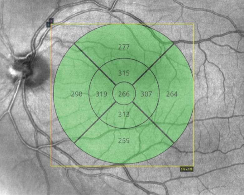
Key content and findings: Unlike the first-generation time-domain OCT (TD-OCT) instruments, the newer spectral-domain OCT (SD-OCT) instruments use a broadband light source to increase axial image resolution. In addition, the decreased image acquisition time also increases the transverse image resolution, reduces motion artifacts, and allows serial cross-sectional images of the retina to be obtained rapidly. A three-dimensional (3D) image of the retina, reconstructed using serial two-dimensional (2D) OCT images, can be used to quantitate retinal thickness and volume and perform analysis of retinal topography. Currently, commercial SD-OCT instruments are used routinely in clinical practice to obtain morphologic information used to diagnose and manage patients with various retinal disorders including macular degeneration and diabetic retinopathy. Newer swept-source OCT technology with faster image acquisition, provides wider field imaging of the peripheral retina. SD-OCT instruments can be incorporated into surgical microscopes to allow imaging of the retina during retinal surgery so that morphologic changes in the retina from surgical maneuvers can be observed in real time. More recently, OCT angiography (OCTA) has been developed which allows rapid, non-invasive 3D imaging of retinal and choroidal vascular flow. This is achieved by processing rapid serial SD-OCT images to detect movement of blood cells within vessels. Research has been done to further improve image resolution of SD-OCT to a cellular level by adding adaptive optics (AO) technology. The latest in SD-OCT technology is optoretinography (ORG), a technique to derive functional information from OCT images of the retina.
Conclusions: Advancement in OCT technology has made it possible to obtain high resolution retinal images that can provide anatomic, physiologic and functional information of the retina in live patients.
期刊介绍:
The Annals of Translational Medicine (Ann Transl Med; ATM; Print ISSN 2305-5839; Online ISSN 2305-5847) is an international, peer-reviewed Open Access journal featuring original and observational investigations in the broad fields of laboratory, clinical, and public health research, aiming to provide practical up-to-date information in significant research from all subspecialties of medicine and to broaden the readers’ vision and horizon from bench to bed and bed to bench. It is published quarterly (April 2013- Dec. 2013), monthly (Jan. 2014 - Feb. 2015), biweekly (March 2015-) and openly distributed worldwide. Annals of Translational Medicine is indexed in PubMed in Sept 2014 and in SCIE in 2018. Specific areas of interest include, but not limited to, multimodality therapy, epidemiology, biomarkers, imaging, biology, pathology, and technical advances related to medicine. Submissions describing preclinical research with potential for application to human disease, and studies describing research obtained from preliminary human experimentation with potential to further the understanding of biological mechanism underlying disease are encouraged. Also warmly welcome are studies describing public health research pertinent to clinic, disease diagnosis and prevention, or healthcare policy. With a focus on interdisciplinary academic cooperation, ATM aims to expedite the translation of scientific discovery into new or improved standards of management and health outcomes practice.
 求助内容:
求助内容: 应助结果提醒方式:


