{"title":"Potential of C-X-C-Chemokine-Receptor-Type-4-Directed PET/CT Using [¹⁸F]AlF-NOTA-QHY-04 in Identifying Molecular Subtypes of Small Cell Lung Cancer.","authors":"Yuxi Luo, Kai Cheng, Jingru Liu, Jinli Pei, Shengnan Xu, Xinzhi Zhao, Shijie Wang, Kunlong Zhao, Wanhu Li, Jie Liu, Jinming Yu","doi":"10.3348/kjr.2024.1266","DOIUrl":null,"url":null,"abstract":"<p><strong>Objective: </strong>Molecular subtyping of small-cell lung cancer (SCLC) has major implications for prognostic relevance and treatment guidance. This study aimed to explore the feasibility of a novel tracer targeting C-X-C-chemokine-receptor-type-4 (CXCR4) for distinguishing different SCLC subtypes.</p><p><strong>Materials and methods: </strong>Thirty-five patients with pathologically confirmed SCLC were enrolled in this prospective study. Immunohistochemical staining was performed to classify the molecular subtypes into SCLC-A, SCLC-N, SCLC-P, and SCLC-I. [¹⁸F]AlF-NOTA-QHY-04 PET/CT parameters were obtained, including the maximum, mean, and peak standard uptake values (SUV<sub>max</sub>, SUV<sub>mean</sub>, and SUV<sub>peak</sub>, respectively) and the ratios of tumors (T) and normal tissues (NT) based on the SUV<sub>max</sub> (T/NT). These parameters were compared among the molecular subtypes. A receiver operating characteristic (ROC) curve was used to analyze the performance of the parameters for distinguishing SCLC-N from other subtypes and neuroendocrine (NE) subtypes (SCLC-A and SCLC-N) from non-NE subtypes (SCLC-P and SCLC-I).</p><p><strong>Results: </strong>The molecular subtypes were SCLC-A (n = 17), SCLC-N (n = 6), SCLC-P (n = 7), and SCLC-I (n = 5). The SCLC-N subtype exhibited significantly higher uptake in both primary tumors and lymph node metastases than the other three subtypes (<i>P</i> < 0.05). When SCLC-N was compared with the other three subtypes combined (referred to as \"other SCLCs\"), all parameters were significantly higher in the SCLC-N group (<i>P</i> < 0.05). ROC analysis showed that these parameters had high accuracy in distinguishing SCLC-N from other SCLCs (area under ROC curve: 0.868-0.948 for primary tumors and 0.783-0.888 for lymph node metastases). Compared with the non-NE group, the SUV<sub>max</sub>, SUV<sub>mean</sub>, and T/NT<sub>lung</sub> were significantly higher in the NE group for primary tumors. ROC analysis showed moderate accuracy in distinguishing between the NE and non-NE groups (ROC area: 0.692-0.786 for primary tumors and 0.692-0.815 for lymph node metastases).</p><p><strong>Conclusion: </strong>Our preliminary findings indicate that CXCR4-directed PET/CT imaging using [¹⁸F]AlF-NOTA-QHY-04 may differentiate between SCLC-N and other molecular subtypes and between NE and non-NE subtypes of SCLC.</p>","PeriodicalId":17881,"journal":{"name":"Korean Journal of Radiology","volume":"26 6","pages":"593-603"},"PeriodicalIF":5.3000,"publicationDate":"2025-06-01","publicationTypes":"Journal Article","fieldsOfStudy":null,"isOpenAccess":false,"openAccessPdf":"https://www.ncbi.nlm.nih.gov/pmc/articles/PMC12123077/pdf/","citationCount":"0","resultStr":null,"platform":"Semanticscholar","paperid":null,"PeriodicalName":"Korean Journal of Radiology","FirstCategoryId":"3","ListUrlMain":"https://doi.org/10.3348/kjr.2024.1266","RegionNum":2,"RegionCategory":"医学","ArticlePicture":[],"TitleCN":null,"AbstractTextCN":null,"PMCID":null,"EPubDate":"","PubModel":"","JCR":"Q1","JCRName":"RADIOLOGY, NUCLEAR MEDICINE & MEDICAL IMAGING","Score":null,"Total":0}
引用次数: 0
Abstract
Objective: Molecular subtyping of small-cell lung cancer (SCLC) has major implications for prognostic relevance and treatment guidance. This study aimed to explore the feasibility of a novel tracer targeting C-X-C-chemokine-receptor-type-4 (CXCR4) for distinguishing different SCLC subtypes.
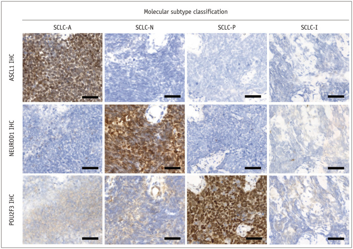
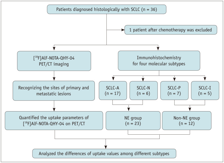
Materials and methods: Thirty-five patients with pathologically confirmed SCLC were enrolled in this prospective study. Immunohistochemical staining was performed to classify the molecular subtypes into SCLC-A, SCLC-N, SCLC-P, and SCLC-I. [¹⁸F]AlF-NOTA-QHY-04 PET/CT parameters were obtained, including the maximum, mean, and peak standard uptake values (SUVmax, SUVmean, and SUVpeak, respectively) and the ratios of tumors (T) and normal tissues (NT) based on the SUVmax (T/NT). These parameters were compared among the molecular subtypes. A receiver operating characteristic (ROC) curve was used to analyze the performance of the parameters for distinguishing SCLC-N from other subtypes and neuroendocrine (NE) subtypes (SCLC-A and SCLC-N) from non-NE subtypes (SCLC-P and SCLC-I).
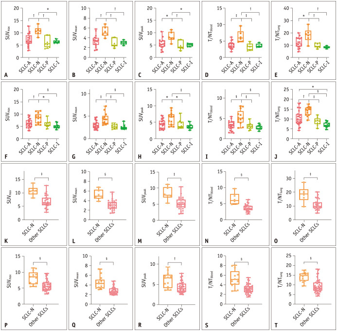
Results: The molecular subtypes were SCLC-A (n = 17), SCLC-N (n = 6), SCLC-P (n = 7), and SCLC-I (n = 5). The SCLC-N subtype exhibited significantly higher uptake in both primary tumors and lymph node metastases than the other three subtypes (P < 0.05). When SCLC-N was compared with the other three subtypes combined (referred to as "other SCLCs"), all parameters were significantly higher in the SCLC-N group (P < 0.05). ROC analysis showed that these parameters had high accuracy in distinguishing SCLC-N from other SCLCs (area under ROC curve: 0.868-0.948 for primary tumors and 0.783-0.888 for lymph node metastases). Compared with the non-NE group, the SUVmax, SUVmean, and T/NTlung were significantly higher in the NE group for primary tumors. ROC analysis showed moderate accuracy in distinguishing between the NE and non-NE groups (ROC area: 0.692-0.786 for primary tumors and 0.692-0.815 for lymph node metastases).
Conclusion: Our preliminary findings indicate that CXCR4-directed PET/CT imaging using [¹⁸F]AlF-NOTA-QHY-04 may differentiate between SCLC-N and other molecular subtypes and between NE and non-NE subtypes of SCLC.
期刊介绍:
The inaugural issue of the Korean J Radiol came out in March 2000. Our journal aims to produce and propagate knowledge on radiologic imaging and related sciences.
A unique feature of the articles published in the Journal will be their reflection of global trends in radiology combined with an East-Asian perspective. Geographic differences in disease prevalence will be reflected in the contents of papers, and this will serve to enrich our body of knowledge.
World''s outstanding radiologists from many countries are serving as editorial board of our journal.

 求助内容:
求助内容: 应助结果提醒方式:


