{"title":"A Nomogram Incorporating Intermuscular Adipose Tissue to Predict Chemotherapy Toxicity in Older Adults With Early-Stage Breast Cancer.","authors":"Wen-Juan Huang, Han-Bing Xie, Lin Zhao, Rui-Han Zhou, Shurui Wang, Xin Zhang, Rui-Tao Wang, Zexun Duan","doi":"10.4048/jbc.2024.0194","DOIUrl":null,"url":null,"abstract":"<p><strong>Purpose: </strong>Recent studies have shown that intermuscular adipose tissue (IMAT) is a significant prognostic factor for breast cancer. To date, no clinical studies have investigated whether IMAT can be used to predict chemotherapy toxicity in older adult patients with early-stage breast cancer.</p><p><strong>Methods: </strong>We included 304 patients diagnosed with stage I-III breast cancer between January 2020 and December 2022 in Harbin Medical University Cancer Hospital. All patients were aged ≥ 65 years and treated with neoadjuvant or adjuvant chemotherapy. IMAT within the pectoralis muscle was measured using computed tomography imaging. Logistic regression analysis was used to identify independent predictors of chemotherapy toxicity. A nomogram was built, and the model performance was assessed using accuracy, discrimination, and clinical benefits. The net reclassification index (NRI) and integrated discrimination improvement (IDI) were used to evaluate changes in model performance after the addition of adipose tissue.</p><p><strong>Results: </strong>Of the 304 patients (184 in the training cohort and 120 in the validation cohort), 30.3% developed grade 3-5 chemotherapy toxicities. Three independent predictors were identified in the multivariate analysis: hemoglobin level, IMAT area, and primary prophylaxis with granulocyte colony-stimulating factor. The nomogram demonstrated area under the receiver operating characteristic curve values of 0.708 (95% confidence interval [CI], 0.616-0.801) and 0.751 (95% CI, 0.655-0.846) in the training and validation cohorts, respectively. The nomogram showed good calibration (Hosmer-Lemeshow test, <i>p</i> > 0.05), and incorporating IMAT improved nomogram performance in both cohorts (all NRI and IDI > 0, <i>p</i> < 0.05). Decision curve analysis revealed that the nomogram was clinically useful.</p><p><strong>Conclusion: </strong>A nomogram including IMAT may be useful for predicting the individual probability of chemotherapy toxicity and guiding therapy in older adults with early-stage breast cancer.</p>","PeriodicalId":15206,"journal":{"name":"Journal of Breast Cancer","volume":" ","pages":"125-138"},"PeriodicalIF":2.4000,"publicationDate":"2025-06-01","publicationTypes":"Journal Article","fieldsOfStudy":null,"isOpenAccess":false,"openAccessPdf":"https://www.ncbi.nlm.nih.gov/pmc/articles/PMC12230286/pdf/","citationCount":"0","resultStr":null,"platform":"Semanticscholar","paperid":null,"PeriodicalName":"Journal of Breast Cancer","FirstCategoryId":"3","ListUrlMain":"https://doi.org/10.4048/jbc.2024.0194","RegionNum":4,"RegionCategory":"医学","ArticlePicture":[],"TitleCN":null,"AbstractTextCN":null,"PMCID":null,"EPubDate":"2025/5/12 0:00:00","PubModel":"Epub","JCR":"Q3","JCRName":"ONCOLOGY","Score":null,"Total":0}
引用次数: 0
Abstract
Purpose: Recent studies have shown that intermuscular adipose tissue (IMAT) is a significant prognostic factor for breast cancer. To date, no clinical studies have investigated whether IMAT can be used to predict chemotherapy toxicity in older adult patients with early-stage breast cancer.
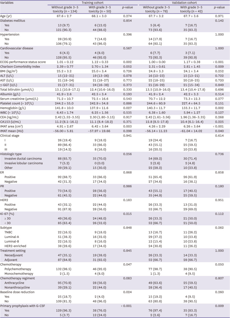
Methods: We included 304 patients diagnosed with stage I-III breast cancer between January 2020 and December 2022 in Harbin Medical University Cancer Hospital. All patients were aged ≥ 65 years and treated with neoadjuvant or adjuvant chemotherapy. IMAT within the pectoralis muscle was measured using computed tomography imaging. Logistic regression analysis was used to identify independent predictors of chemotherapy toxicity. A nomogram was built, and the model performance was assessed using accuracy, discrimination, and clinical benefits. The net reclassification index (NRI) and integrated discrimination improvement (IDI) were used to evaluate changes in model performance after the addition of adipose tissue.
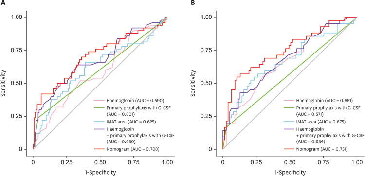
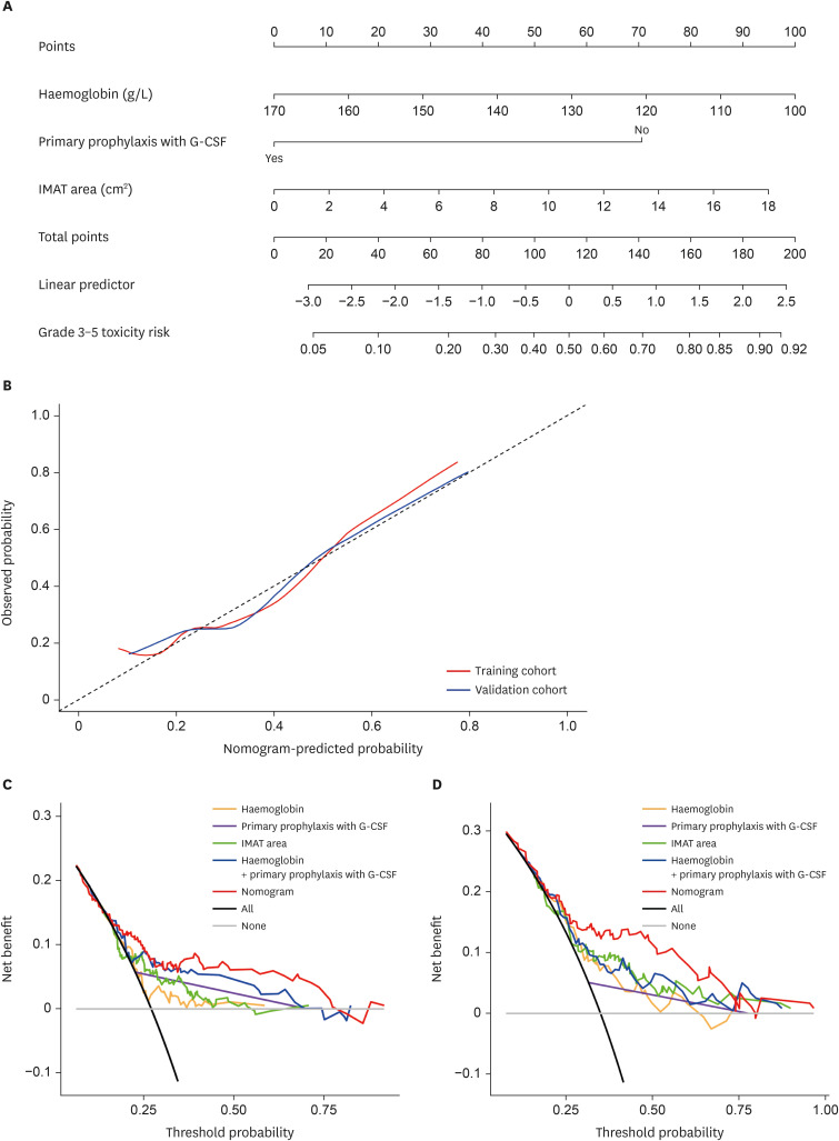
Results: Of the 304 patients (184 in the training cohort and 120 in the validation cohort), 30.3% developed grade 3-5 chemotherapy toxicities. Three independent predictors were identified in the multivariate analysis: hemoglobin level, IMAT area, and primary prophylaxis with granulocyte colony-stimulating factor. The nomogram demonstrated area under the receiver operating characteristic curve values of 0.708 (95% confidence interval [CI], 0.616-0.801) and 0.751 (95% CI, 0.655-0.846) in the training and validation cohorts, respectively. The nomogram showed good calibration (Hosmer-Lemeshow test, p > 0.05), and incorporating IMAT improved nomogram performance in both cohorts (all NRI and IDI > 0, p < 0.05). Decision curve analysis revealed that the nomogram was clinically useful.
Conclusion: A nomogram including IMAT may be useful for predicting the individual probability of chemotherapy toxicity and guiding therapy in older adults with early-stage breast cancer.
期刊介绍:
The Journal of Breast Cancer (abbreviated as ''J Breast Cancer'') is the official journal of the Korean Breast Cancer Society, which is issued quarterly in the last day of March, June, September, and December each year since 1998. All the contents of the Journal is available online at the official journal website (http://ejbc.kr) under open access policy. The journal aims to provide a forum for the academic communication between medical doctors, basic science researchers, and health care professionals to be interested in breast cancer. To get this aim, we publish original investigations, review articles, brief communications including case reports, editorial opinions on the topics of importance to breast cancer, and welcome new research findings and epidemiological studies, especially when they contain a regional data to grab the international reader''s interest. Although the journal is mainly dealing with the issues of breast cancer, rare cases among benign breast diseases or evidence-based scientifically written articles providing useful information for clinical practice can be published as well.
 求助内容:
求助内容: 应助结果提醒方式:


