Amelia D Dahlén, Sahar Roshanbin, Ximena Aguilar, Nadja M Bucher, Sara Lopes van den Broek, Dag Sehlin, Stina Syvänen
{"title":"PET imaging of TREM2 in amyloid-beta induced neuroinflammation.","authors":"Amelia D Dahlén, Sahar Roshanbin, Ximena Aguilar, Nadja M Bucher, Sara Lopes van den Broek, Dag Sehlin, Stina Syvänen","doi":"10.1007/s00259-025-07358-0","DOIUrl":null,"url":null,"abstract":"<p><strong>Purpose: </strong>The triggering receptor expressed on myeloid cells 2 (TREM2) has become a promising target for biologics in both monitoring and treating neuroinflammation in Alzheimer's disease (AD). This study aimed to develop and compare bispecific anti-TREM2 antibodies featuring different transferrin receptor (TfR) binders to enhance brain delivery, identifying the most suitable format for in vivo PET imaging of TREM2 in transgenic AD mice.</p><p><strong>Methods: </strong>Three bispecific TREM2-antibody formats were produced and evaluated for their ability to cross the blood-brain barrier (BBB) via TfR-mediated transcytosis and bind TREM2. Blood concentration profiles up to 72 h post-injection (p.i.), and ex vivo brain uptake of iodine-125-labeled antibody constructs were quantified in App<sup>NL-G-F</sup> and age-matched wild type (WT) mice using a γ-counter. The best-performing bispecific TREM2-antibody was radiolabeled with iodine-124 and used for in vivo PET imaging of brain TREM2 levels in App<sup>NL-G-F</sup> mice at 72 h p.i. Brain TREM2 concentrations were subsequently quantified using ELISA.</p><p><strong>Results: </strong>The antibody format carrying two scFv8D3 TfR-binders (IgG-scFv<sub>2</sub>), demonstrated the highest brain concentrations of all tested bispecific constructs. This antibody also exhibited significantly higher brain concentrations in App<sup>NL-G-F</sup> mice compared to WT mice at both 48 and 72 h p.i. This difference was further visualized and quantified through in vivo PET imaging. Moreover, brain concentrations of the antibody ligand correlated with elevated TREM2 levels in brain homogenates.</p><p><strong>Conclusion: </strong>These findings highlight IgG-scFv<sub>2</sub> as a promising radioligand for in vivo PET imaging of TREM2, advancing non-invasive neuroinflammation studies and supporting drug development for AD and other neurodegenerative diseases.</p>","PeriodicalId":11909,"journal":{"name":"European Journal of Nuclear Medicine and Molecular Imaging","volume":" ","pages":"4320-4333"},"PeriodicalIF":7.6000,"publicationDate":"2025-09-01","publicationTypes":"Journal Article","fieldsOfStudy":null,"isOpenAccess":false,"openAccessPdf":"https://www.ncbi.nlm.nih.gov/pmc/articles/PMC12396982/pdf/","citationCount":"0","resultStr":null,"platform":"Semanticscholar","paperid":null,"PeriodicalName":"European Journal of Nuclear Medicine and Molecular Imaging","FirstCategoryId":"3","ListUrlMain":"https://doi.org/10.1007/s00259-025-07358-0","RegionNum":1,"RegionCategory":"医学","ArticlePicture":[],"TitleCN":null,"AbstractTextCN":null,"PMCID":null,"EPubDate":"2025/5/28 0:00:00","PubModel":"Epub","JCR":"Q1","JCRName":"RADIOLOGY, NUCLEAR MEDICINE & MEDICAL IMAGING","Score":null,"Total":0}
引用次数: 0
Abstract
Purpose: The triggering receptor expressed on myeloid cells 2 (TREM2) has become a promising target for biologics in both monitoring and treating neuroinflammation in Alzheimer's disease (AD). This study aimed to develop and compare bispecific anti-TREM2 antibodies featuring different transferrin receptor (TfR) binders to enhance brain delivery, identifying the most suitable format for in vivo PET imaging of TREM2 in transgenic AD mice.
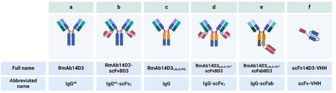
Methods: Three bispecific TREM2-antibody formats were produced and evaluated for their ability to cross the blood-brain barrier (BBB) via TfR-mediated transcytosis and bind TREM2. Blood concentration profiles up to 72 h post-injection (p.i.), and ex vivo brain uptake of iodine-125-labeled antibody constructs were quantified in AppNL-G-F and age-matched wild type (WT) mice using a γ-counter. The best-performing bispecific TREM2-antibody was radiolabeled with iodine-124 and used for in vivo PET imaging of brain TREM2 levels in AppNL-G-F mice at 72 h p.i. Brain TREM2 concentrations were subsequently quantified using ELISA.
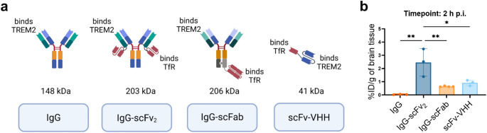
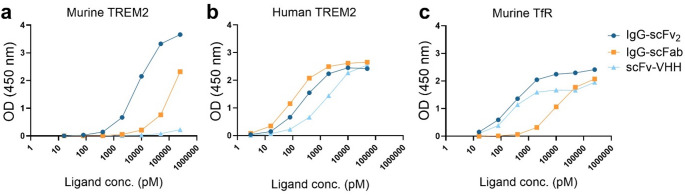
Results: The antibody format carrying two scFv8D3 TfR-binders (IgG-scFv2), demonstrated the highest brain concentrations of all tested bispecific constructs. This antibody also exhibited significantly higher brain concentrations in AppNL-G-F mice compared to WT mice at both 48 and 72 h p.i. This difference was further visualized and quantified through in vivo PET imaging. Moreover, brain concentrations of the antibody ligand correlated with elevated TREM2 levels in brain homogenates.
Conclusion: These findings highlight IgG-scFv2 as a promising radioligand for in vivo PET imaging of TREM2, advancing non-invasive neuroinflammation studies and supporting drug development for AD and other neurodegenerative diseases.
期刊介绍:
The European Journal of Nuclear Medicine and Molecular Imaging serves as a platform for the exchange of clinical and scientific information within nuclear medicine and related professions. It welcomes international submissions from professionals involved in the functional, metabolic, and molecular investigation of diseases. The journal's coverage spans physics, dosimetry, radiation biology, radiochemistry, and pharmacy, providing high-quality peer review by experts in the field. Known for highly cited and downloaded articles, it ensures global visibility for research work and is part of the EJNMMI journal family.

 求助内容:
求助内容: 应助结果提醒方式:


