Sliman Marina, Sham Zoukar, Mais Basel Alreem Mohaisen, Miriam Laflouf, Mahmoud Alhamadeh Alswij, Ali Al Wynse, Alhamza Khalaf Alali, Mouhammed Sleiay
{"title":"A Rare Case Report of Pancreatic Hydatid Cyst From Syria: A Diagnostic and Therapeutic Challenge.","authors":"Sliman Marina, Sham Zoukar, Mais Basel Alreem Mohaisen, Miriam Laflouf, Mahmoud Alhamadeh Alswij, Ali Al Wynse, Alhamza Khalaf Alali, Mouhammed Sleiay","doi":"10.1177/11795476251342378","DOIUrl":null,"url":null,"abstract":"<p><strong>Introduction and significance: </strong>Hydatid disease, primarily caused by the parasite <i>Echinococcus granulosus</i>, commonly affects the liver and lungs. However, it can also involve other organs, including the pancreas. Pancreatic hydatid cysts are rare, constituting less than 2% of all hydatid cases. Their infrequent occurrence and atypical presentation often pose diagnostic and therapeutic challenges, especially in non-endemic regions.</p><p><strong>Case presentation: </strong>A 42-year-old man with a cystic lesion in his pancreatic tail who had no notable medical history was seen. The diagnosis of a pancreatic hydatid cyst was validated by serological testing and diagnostic imaging techniques.</p><p><strong>Clinical discussion: </strong>Due to their uncommon nature, the differential diagnosis of pancreatic hydatid cysts can be challenging. Imaging modalities such as computed tomography (CT), magnetic resonance imaging (MRI), and ultrasound are important for identifying characteristic features. Serological testing further aids in confirming the diagnosis. Treatment typically involves a combination of medical and surgical approaches. Antiparasitic drugs, such as albendazole or mebendazole, are administered to kill the parasite. Surgical intervention is necessary to remove the cyst and reduce the chance of recurrence and complications.</p><p><strong>Conclusion: </strong>This instance highlights the significance it is to take hydatid disease into consideration when making a differential diagnosis for pancreatic cystic lesions, particularly in people from endemic regions. For the optimal possible patient outcomes and avoiding of complications, early diagnosis and effective treatment are important.</p>","PeriodicalId":10357,"journal":{"name":"Clinical Medicine Insights. Case Reports","volume":"18 ","pages":"11795476251342378"},"PeriodicalIF":0.6000,"publicationDate":"2025-05-26","publicationTypes":"Journal Article","fieldsOfStudy":null,"isOpenAccess":false,"openAccessPdf":"https://www.ncbi.nlm.nih.gov/pmc/articles/PMC12107011/pdf/","citationCount":"0","resultStr":null,"platform":"Semanticscholar","paperid":null,"PeriodicalName":"Clinical Medicine Insights. Case Reports","FirstCategoryId":"1085","ListUrlMain":"https://doi.org/10.1177/11795476251342378","RegionNum":0,"RegionCategory":null,"ArticlePicture":[],"TitleCN":null,"AbstractTextCN":null,"PMCID":null,"EPubDate":"2025/1/1 0:00:00","PubModel":"eCollection","JCR":"Q3","JCRName":"MEDICINE, GENERAL & INTERNAL","Score":null,"Total":0}
引用次数: 0
Abstract
Introduction and significance: Hydatid disease, primarily caused by the parasite Echinococcus granulosus, commonly affects the liver and lungs. However, it can also involve other organs, including the pancreas. Pancreatic hydatid cysts are rare, constituting less than 2% of all hydatid cases. Their infrequent occurrence and atypical presentation often pose diagnostic and therapeutic challenges, especially in non-endemic regions.
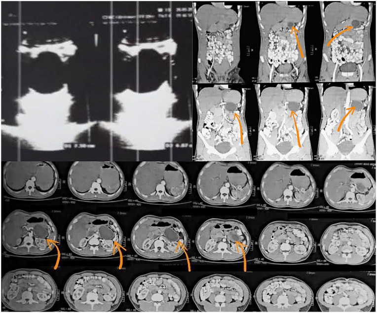
Case presentation: A 42-year-old man with a cystic lesion in his pancreatic tail who had no notable medical history was seen. The diagnosis of a pancreatic hydatid cyst was validated by serological testing and diagnostic imaging techniques.
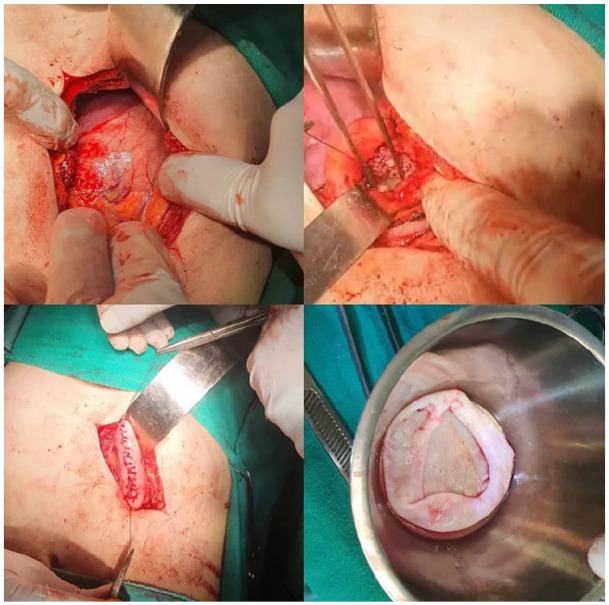
Clinical discussion: Due to their uncommon nature, the differential diagnosis of pancreatic hydatid cysts can be challenging. Imaging modalities such as computed tomography (CT), magnetic resonance imaging (MRI), and ultrasound are important for identifying characteristic features. Serological testing further aids in confirming the diagnosis. Treatment typically involves a combination of medical and surgical approaches. Antiparasitic drugs, such as albendazole or mebendazole, are administered to kill the parasite. Surgical intervention is necessary to remove the cyst and reduce the chance of recurrence and complications.
Conclusion: This instance highlights the significance it is to take hydatid disease into consideration when making a differential diagnosis for pancreatic cystic lesions, particularly in people from endemic regions. For the optimal possible patient outcomes and avoiding of complications, early diagnosis and effective treatment are important.

 求助内容:
求助内容: 应助结果提醒方式:


