Mesenchymal stem cells reduce the genotoxic effect of lead acetate in the testis of male rats and induce testicular cellular proliferation indicated by 16S rRNA sequence, increase the proliferation marker Ki-67 and a reduction in the apoptosis marker caspase-3.
Mohamed Allam, Yahia A Amin, Samer S Fouad, Rana A Ali, Mariam A Fawy, Maha Abd-El Baki Ahmed, Rana Toghan, Lobna A Ali
{"title":"Mesenchymal stem cells reduce the genotoxic effect of lead acetate in the testis of male rats and induce testicular cellular proliferation indicated by 16S rRNA sequence, increase the proliferation marker Ki-67 and a reduction in the apoptosis marker caspase-3.","authors":"Mohamed Allam, Yahia A Amin, Samer S Fouad, Rana A Ali, Mariam A Fawy, Maha Abd-El Baki Ahmed, Rana Toghan, Lobna A Ali","doi":"10.1186/s40659-025-00614-5","DOIUrl":null,"url":null,"abstract":"<p><strong>Background: </strong>Lead is a ubiquitous environmental and industrial pollutant with worldwide health problems. Lead acetate toxicity induces both genotoxic effects and apoptosis. The present study aimed to investigate the usage of mesenchymal stem cells (MSCs) in the treatment of the genotoxic effect of lead acetate (LA) in the testis and its effect on the expression of the apoptosis marker caspase-3 and the proliferation marker Ki-67 in the injured testicular tissue.</p><p><strong>Methods: </strong>Twenty-one adult male rats were used in this investigation (7 rats/group). Group I received saline and served as the control group (ctrl group); Group II received lead acetate (100 mg/kg) and was called the LA group; Group III received both lead acetate (100 mg/kg) and MSCs (1 × 10<sup>6</sup> cells/rat) and was called the LA-MSCs group. Body and testis weight, plus semen analysis, were performed in all groups. Reproductive hormones, serotonin, and cortisol were determined in sera. Additionally, oxidative/antioxidative status and lead acetate-induced genetic variations were investigated. Immunohistochemical staining for the proliferation marker Ki-67 and the apoptosis marker caspase-3 was also performed.</p><p><strong>Results: </strong>revealed that the weight of the body and testis and semen parameters (sperm count, viability, and motility) of the LA group exhibited significant reduction compared to the Ctrl and the LA-MSCs group. In addition, the LA group showed reproductive hormonal imbalance and an increase in oxidative stress biomarkers compared to the LA-MSCs group that showed a significant improvement in these parameters. Compared to the ctrl group, the LA group showed a highly genetic distance value (0.0031), while the LA-MSCs group showed a low genetic distance value (0.0019). This illustrated that the LA-MSCs group exhibited reduced genetic variation induced by LA compared to the LA group. Histological evaluation indicated the presence of severe diffuse degeneration and necrosis in the spermatocytes in the LA group compared to the control one, while co-treatment by MSCs induced significant reduction in these degenerative changes. Immunohistochemical investigation revealed increased expression of the caspase-3 antibody in the testicular tissue of the LA group, while it is significantly decreased in the LA- MSCs group. In contrast, the KI67 antibody revealed a significant decrease in its expression in the LA group, while it was significantly increased in the LA-MSCs group after treatment by MSCs.</p><p><strong>Conclusions: </strong>It can be concluded that the MSCs are a potential therapeutic for the treatment of testicular dysfunction induced by LA through the reduction of oxidative stress, genotoxic effect, and apoptosis marker caspase-3, and an increase in the proliferation marker Ki-67 in the testicular tissue associated with restoration of hormonal imbalance.</p>","PeriodicalId":9084,"journal":{"name":"Biological Research","volume":"58 1","pages":"29"},"PeriodicalIF":4.6000,"publicationDate":"2025-05-27","publicationTypes":"Journal Article","fieldsOfStudy":null,"isOpenAccess":false,"openAccessPdf":"https://www.ncbi.nlm.nih.gov/pmc/articles/PMC12117784/pdf/","citationCount":"0","resultStr":null,"platform":"Semanticscholar","paperid":null,"PeriodicalName":"Biological Research","FirstCategoryId":"99","ListUrlMain":"https://doi.org/10.1186/s40659-025-00614-5","RegionNum":2,"RegionCategory":"生物学","ArticlePicture":[],"TitleCN":null,"AbstractTextCN":null,"PMCID":null,"EPubDate":"","PubModel":"","JCR":"Q1","JCRName":"BIOLOGY","Score":null,"Total":0}
引用次数: 0
Abstract
Background: Lead is a ubiquitous environmental and industrial pollutant with worldwide health problems. Lead acetate toxicity induces both genotoxic effects and apoptosis. The present study aimed to investigate the usage of mesenchymal stem cells (MSCs) in the treatment of the genotoxic effect of lead acetate (LA) in the testis and its effect on the expression of the apoptosis marker caspase-3 and the proliferation marker Ki-67 in the injured testicular tissue.
Methods: Twenty-one adult male rats were used in this investigation (7 rats/group). Group I received saline and served as the control group (ctrl group); Group II received lead acetate (100 mg/kg) and was called the LA group; Group III received both lead acetate (100 mg/kg) and MSCs (1 × 10⁶ cells/rat) and was called the LA-MSCs group. Body and testis weight, plus semen analysis, were performed in all groups. Reproductive hormones, serotonin, and cortisol were determined in sera. Additionally, oxidative/antioxidative status and lead acetate-induced genetic variations were investigated. Immunohistochemical staining for the proliferation marker Ki-67 and the apoptosis marker caspase-3 was also performed.
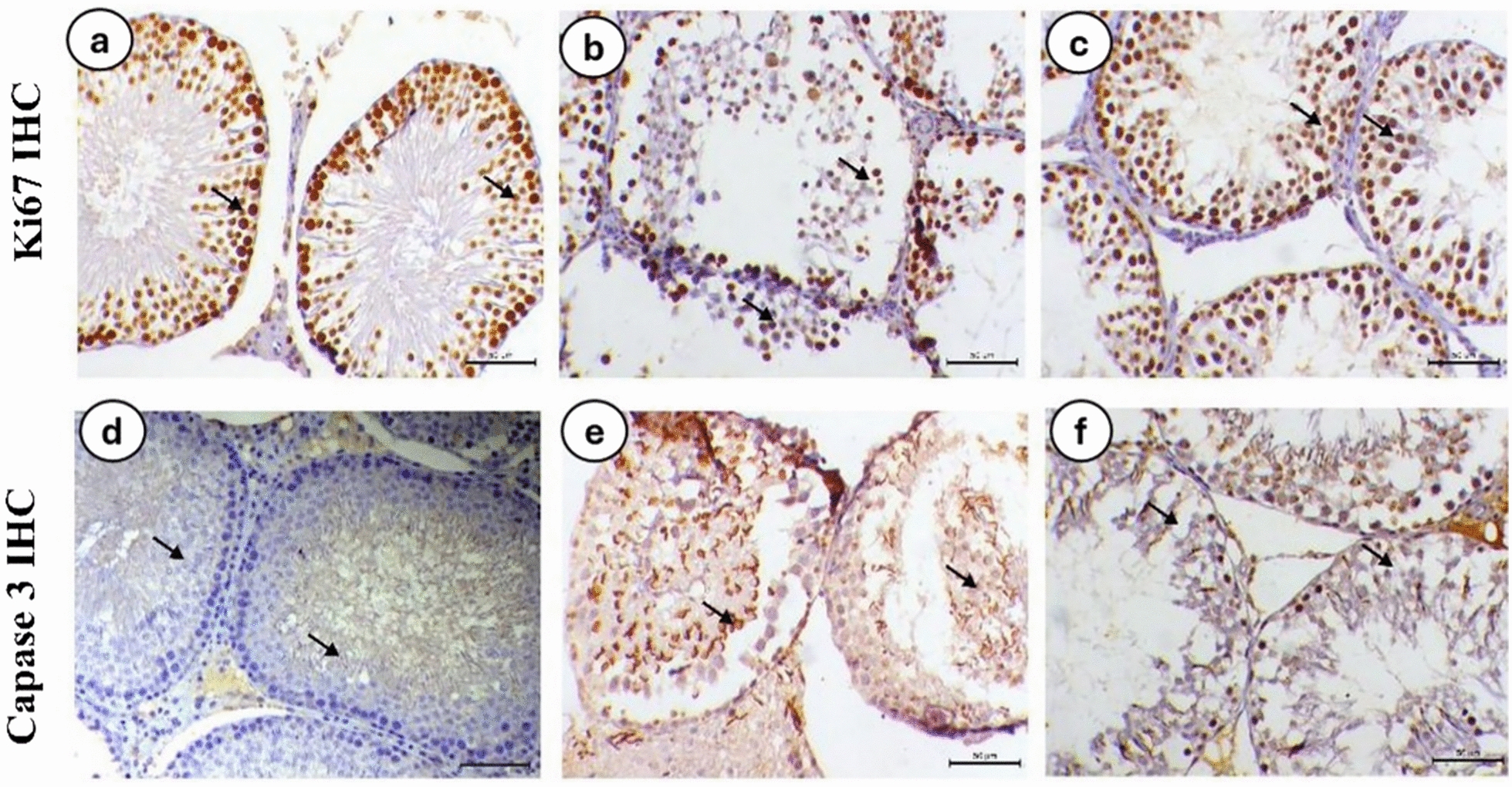
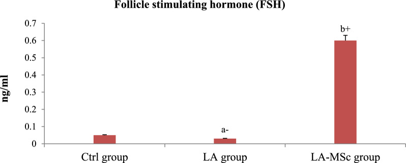
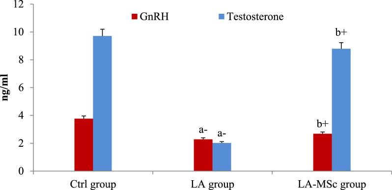
Results: revealed that the weight of the body and testis and semen parameters (sperm count, viability, and motility) of the LA group exhibited significant reduction compared to the Ctrl and the LA-MSCs group. In addition, the LA group showed reproductive hormonal imbalance and an increase in oxidative stress biomarkers compared to the LA-MSCs group that showed a significant improvement in these parameters. Compared to the ctrl group, the LA group showed a highly genetic distance value (0.0031), while the LA-MSCs group showed a low genetic distance value (0.0019). This illustrated that the LA-MSCs group exhibited reduced genetic variation induced by LA compared to the LA group. Histological evaluation indicated the presence of severe diffuse degeneration and necrosis in the spermatocytes in the LA group compared to the control one, while co-treatment by MSCs induced significant reduction in these degenerative changes. Immunohistochemical investigation revealed increased expression of the caspase-3 antibody in the testicular tissue of the LA group, while it is significantly decreased in the LA- MSCs group. In contrast, the KI67 antibody revealed a significant decrease in its expression in the LA group, while it was significantly increased in the LA-MSCs group after treatment by MSCs.
Conclusions: It can be concluded that the MSCs are a potential therapeutic for the treatment of testicular dysfunction induced by LA through the reduction of oxidative stress, genotoxic effect, and apoptosis marker caspase-3, and an increase in the proliferation marker Ki-67 in the testicular tissue associated with restoration of hormonal imbalance.
期刊介绍:
Biological Research is an open access, peer-reviewed journal that encompasses diverse fields of experimental biology, such as biochemistry, bioinformatics, biotechnology, cell biology, cancer, chemical biology, developmental biology, evolutionary biology, genetics, genomics, immunology, marine biology, microbiology, molecular biology, neuroscience, plant biology, physiology, stem cell research, structural biology and systems biology.
 求助内容:
求助内容: 应助结果提醒方式:


