Sharmeen Sorathia, Aro Daniela Arockiam, Ankit Agrawal, Elio Haroun, Rishabh Khurana, Abdelrahman Ahmed, Leslie Cho, Wael Jaber, Brian Griffin, Tom Kai Ming Wang
{"title":"Contemporary review of the clinical features, multi-modality imaging, and management of coronary artery aneurysms.","authors":"Sharmeen Sorathia, Aro Daniela Arockiam, Ankit Agrawal, Elio Haroun, Rishabh Khurana, Abdelrahman Ahmed, Leslie Cho, Wael Jaber, Brian Griffin, Tom Kai Ming Wang","doi":"10.1093/ehjimp/qyaf054","DOIUrl":null,"url":null,"abstract":"<p><p>Coronary artery aneurysm (CAA) is a rare, dilated segment of the coronary artery. Phenotypically, it can have different morphologies, patterns, content, and affected vessels, making multimodality invasive and noninvasive imaging play a significant role in its evaluation. In this review article, we discuss in depth the contemporary perspectives in pathophysiology, epidemiology, clinical presentation, multimodality imaging evaluation, treatment strategies (medical, percutaneous and surgical) and outcomes of CAA. CAA is predominantly associated with coronary atherosclerotic diseases and/or autoimmune vasculitis conditions including Kawasaki disease. It can present with various clinical manifestations, from an incidental finding to myocardial infarction and life-threatening complications. While coronary angiography has been the gold standard to detect CAA, noninvasive modalities including coronary computed tomography have increasingly been utilized given its high resolution for anatomical depiction of the vessels and relations to surrounding structures. We suggest a stepwise approach to the assessment and clinical decision-making in the management of CAA. Pharmacological strategies include risk factor control and secondary prevention. For obstructive ischaemic presenting aneurysms, percutaneous is pursued, while larger or multi-vessel obstructive lesions are typically managed with coronary surgery.</p>","PeriodicalId":94317,"journal":{"name":"European heart journal. Imaging methods and practice","volume":"3 1","pages":"qyaf054"},"PeriodicalIF":0.0000,"publicationDate":"2025-05-09","publicationTypes":"Journal Article","fieldsOfStudy":null,"isOpenAccess":false,"openAccessPdf":"https://www.ncbi.nlm.nih.gov/pmc/articles/PMC12103974/pdf/","citationCount":"0","resultStr":null,"platform":"Semanticscholar","paperid":null,"PeriodicalName":"European heart journal. Imaging methods and practice","FirstCategoryId":"1085","ListUrlMain":"https://doi.org/10.1093/ehjimp/qyaf054","RegionNum":0,"RegionCategory":null,"ArticlePicture":[],"TitleCN":null,"AbstractTextCN":null,"PMCID":null,"EPubDate":"2025/1/1 0:00:00","PubModel":"eCollection","JCR":"","JCRName":"","Score":null,"Total":0}
引用次数: 0
Abstract
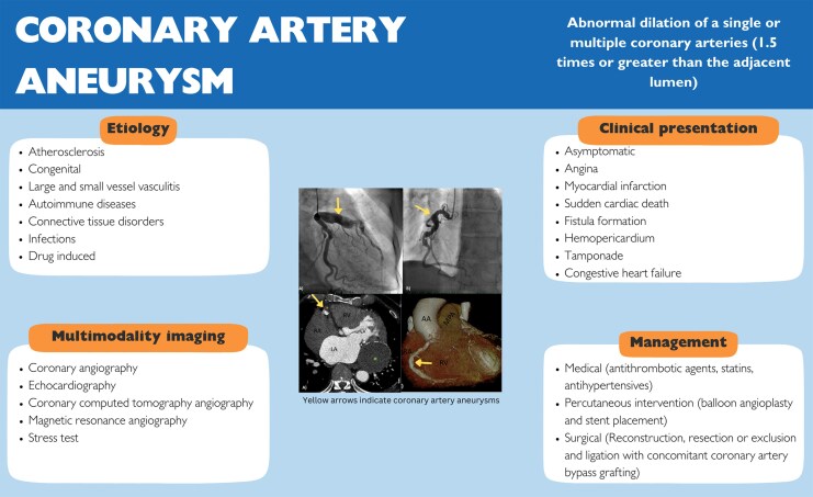
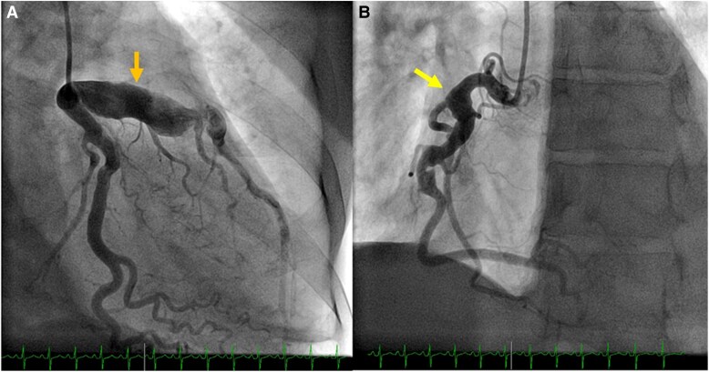
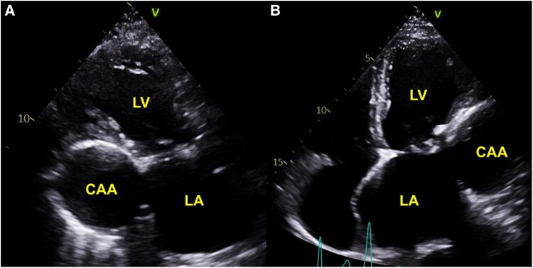
Coronary artery aneurysm (CAA) is a rare, dilated segment of the coronary artery. Phenotypically, it can have different morphologies, patterns, content, and affected vessels, making multimodality invasive and noninvasive imaging play a significant role in its evaluation. In this review article, we discuss in depth the contemporary perspectives in pathophysiology, epidemiology, clinical presentation, multimodality imaging evaluation, treatment strategies (medical, percutaneous and surgical) and outcomes of CAA. CAA is predominantly associated with coronary atherosclerotic diseases and/or autoimmune vasculitis conditions including Kawasaki disease. It can present with various clinical manifestations, from an incidental finding to myocardial infarction and life-threatening complications. While coronary angiography has been the gold standard to detect CAA, noninvasive modalities including coronary computed tomography have increasingly been utilized given its high resolution for anatomical depiction of the vessels and relations to surrounding structures. We suggest a stepwise approach to the assessment and clinical decision-making in the management of CAA. Pharmacological strategies include risk factor control and secondary prevention. For obstructive ischaemic presenting aneurysms, percutaneous is pursued, while larger or multi-vessel obstructive lesions are typically managed with coronary surgery.

 求助内容:
求助内容: 应助结果提醒方式:


