{"title":"Breast Myofibroblastoma: A Single Institutional Case Series.","authors":"Meghna Pinnaka, Melissa Garcia Patino, Vasupriya Ravi, Alia Nazarullah, Ismail Jatoi","doi":"10.4274/ejbh.galenos.2025.2025-3-6","DOIUrl":null,"url":null,"abstract":"<p><strong>Objective: </strong>Breast myofibroblastoma (BM) is a rare, benign mesenchymal tumor primarily affecting older men and postmenopausal women. This study analyzed the clinicopathologic features, immunohistochemical profiles, and treatment outcomes of five BM cases diagnosed at a single institution over a period of 20 years.</p><p><strong>Materials and methods: </strong>A retrospective review was conducted for five patients diagnosed with BM between 1998 and 2024. Data included age, clinical presentation, tumor size, histopathologic findings, immunohistochemical profiles, treatment approaches, and follow-up outcomes.</p><p><strong>Results: </strong>The median age at diagnosis was 68 years, with a mean tumor size of 5.06 cm. Clinical presentation included palpable, painless masses in two patients and an incidental finding in one, while data were unavailable for two cases. Histopathology showed well-circumscribed, unencapsulated tumors composed of spindle cells with admixed adipose tissue and collagen bundles. Immunohistochemically, all tumors were positive for desmin and CD34, with variable smooth muscle actin expression and negative S100 staining. No cases exhibited nuclear beta-catenin staining or 13q14 deletions. All patients underwent surgical excision, with one requiring re-excision due to tumor abutting margins. No recurrences were observed during follow-up (2-18 months).</p><p><strong>Conclusion: </strong>BM is a benign tumor with favorable outcomes following surgical excision. This study underscores the variability in immunohistochemical staining and the importance of distinguishing BM from other spindle cell tumors. Increased numbers of published cases and refining diagnostic markers may be important to improve clinical management and reduce diagnostic uncertainty.</p>","PeriodicalId":93996,"journal":{"name":"European journal of breast health","volume":" ","pages":"211-214"},"PeriodicalIF":1.7000,"publicationDate":"2025-06-20","publicationTypes":"Journal Article","fieldsOfStudy":null,"isOpenAccess":false,"openAccessPdf":"https://www.ncbi.nlm.nih.gov/pmc/articles/PMC12180106/pdf/","citationCount":"0","resultStr":null,"platform":"Semanticscholar","paperid":null,"PeriodicalName":"European journal of breast health","FirstCategoryId":"1085","ListUrlMain":"https://doi.org/10.4274/ejbh.galenos.2025.2025-3-6","RegionNum":0,"RegionCategory":null,"ArticlePicture":[],"TitleCN":null,"AbstractTextCN":null,"PMCID":null,"EPubDate":"2025/5/27 0:00:00","PubModel":"Epub","JCR":"Q4","JCRName":"ONCOLOGY","Score":null,"Total":0}
引用次数: 0
Abstract
Objective: Breast myofibroblastoma (BM) is a rare, benign mesenchymal tumor primarily affecting older men and postmenopausal women. This study analyzed the clinicopathologic features, immunohistochemical profiles, and treatment outcomes of five BM cases diagnosed at a single institution over a period of 20 years.
Materials and methods: A retrospective review was conducted for five patients diagnosed with BM between 1998 and 2024. Data included age, clinical presentation, tumor size, histopathologic findings, immunohistochemical profiles, treatment approaches, and follow-up outcomes.
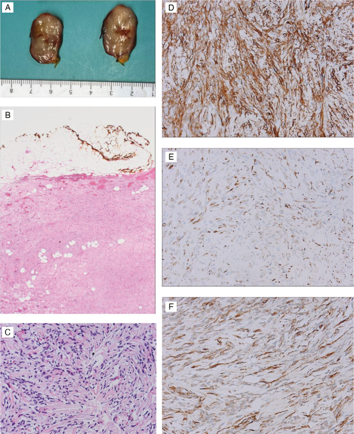
Results: The median age at diagnosis was 68 years, with a mean tumor size of 5.06 cm. Clinical presentation included palpable, painless masses in two patients and an incidental finding in one, while data were unavailable for two cases. Histopathology showed well-circumscribed, unencapsulated tumors composed of spindle cells with admixed adipose tissue and collagen bundles. Immunohistochemically, all tumors were positive for desmin and CD34, with variable smooth muscle actin expression and negative S100 staining. No cases exhibited nuclear beta-catenin staining or 13q14 deletions. All patients underwent surgical excision, with one requiring re-excision due to tumor abutting margins. No recurrences were observed during follow-up (2-18 months).
Conclusion: BM is a benign tumor with favorable outcomes following surgical excision. This study underscores the variability in immunohistochemical staining and the importance of distinguishing BM from other spindle cell tumors. Increased numbers of published cases and refining diagnostic markers may be important to improve clinical management and reduce diagnostic uncertainty.

 求助内容:
求助内容: 应助结果提醒方式:


