Sadie Badrie, Zena Moore, Declan Patton, Tom O'Connor, Rosemarie Derwin
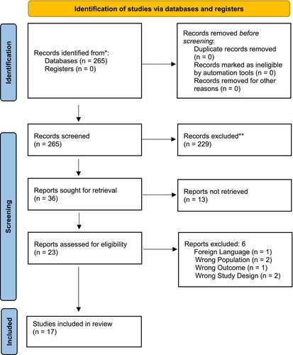
{"title":"The Clinical Utility of Autofluorescence Imaging for Bacterial Detection in Wounds: A Systematic Review","authors":"Sadie Badrie, Zena Moore, Declan Patton, Tom O'Connor, Rosemarie Derwin","doi":"10.1111/iwj.70678","DOIUrl":null,"url":null,"abstract":"<p>This systematic review evaluated the clinical utility and diagnostic accuracy of autofluorescence imaging in detecting bacterial presence in wounds. A literature search was conducted in January 2025 across PubMed, Scopus, Cochrane, and EMBASE databases. Eligible studies included clinical trials and observational studies assessing autofluorescence imaging for wound bacterial detection. Seventeen studies were included; sixteen assessed the MolecuLight i:X device, and one evaluated PRODIGI. Autofluorescence imaging demonstrated higher accuracy than White Light and Clinical Signs and Symptoms-based assessment in detecting bacterial burden. Five studies highlighted its role in enhancing swabbing techniques, with fluorescence-guided sampling yielding higher bacterial counts than conventional methods. Ten studies reported significant bacterial reduction with autofluorescence-guided debridement. Six studies emphasized its role in refining treatment decisions and accelerating wound healing. Quality appraisal was undertaken using Evidence-Based Librarianship criteria, which deemed 10 studies valid, while 7 had limitations related to population representation. In conclusion, autofluorescence imaging enhances wound assessment by improving bacterial detection and may support more targeted clinical interventions. However, further research is needed to clarify its impact on infection control and long-term healing outcomes.</p>","PeriodicalId":14451,"journal":{"name":"International Wound Journal","volume":"22 6","pages":""},"PeriodicalIF":2.5000,"publicationDate":"2025-05-28","publicationTypes":"Journal Article","fieldsOfStudy":null,"isOpenAccess":false,"openAccessPdf":"https://onlinelibrary.wiley.com/doi/epdf/10.1111/iwj.70678","citationCount":"0","resultStr":null,"platform":"Semanticscholar","paperid":null,"PeriodicalName":"International Wound Journal","FirstCategoryId":"3","ListUrlMain":"https://onlinelibrary.wiley.com/doi/10.1111/iwj.70678","RegionNum":3,"RegionCategory":"医学","ArticlePicture":[],"TitleCN":null,"AbstractTextCN":null,"PMCID":null,"EPubDate":"","PubModel":"","JCR":"Q2","JCRName":"DERMATOLOGY","Score":null,"Total":0}
引用次数: 0
Abstract
This systematic review evaluated the clinical utility and diagnostic accuracy of autofluorescence imaging in detecting bacterial presence in wounds. A literature search was conducted in January 2025 across PubMed, Scopus, Cochrane, and EMBASE databases. Eligible studies included clinical trials and observational studies assessing autofluorescence imaging for wound bacterial detection. Seventeen studies were included; sixteen assessed the MolecuLight i:X device, and one evaluated PRODIGI. Autofluorescence imaging demonstrated higher accuracy than White Light and Clinical Signs and Symptoms-based assessment in detecting bacterial burden. Five studies highlighted its role in enhancing swabbing techniques, with fluorescence-guided sampling yielding higher bacterial counts than conventional methods. Ten studies reported significant bacterial reduction with autofluorescence-guided debridement. Six studies emphasized its role in refining treatment decisions and accelerating wound healing. Quality appraisal was undertaken using Evidence-Based Librarianship criteria, which deemed 10 studies valid, while 7 had limitations related to population representation. In conclusion, autofluorescence imaging enhances wound assessment by improving bacterial detection and may support more targeted clinical interventions. However, further research is needed to clarify its impact on infection control and long-term healing outcomes.
期刊介绍:
The Editors welcome papers on all aspects of prevention and treatment of wounds and associated conditions in the fields of surgery, dermatology, oncology, nursing, radiotherapy, physical therapy, occupational therapy and podiatry. The Journal accepts papers in the following categories:
- Research papers
- Review articles
- Clinical studies
- Letters
- News and Views: international perspectives, education initiatives, guidelines and different activities of groups and societies.
Calendar of events
The Editors are supported by a board of international experts and a panel of reviewers across a range of disciplines and specialties which ensures only the most current and relevant research is published.
 求助内容:
求助内容: 应助结果提醒方式:


