L Z Sirotina, A L Potapov, M M Loginova, L V Shkalova, A D Varnavskaya, K V Kazakova, A A Chaikin, D A Bolshakova, M A Sirotkina, T M Motovilova, N D Gladkova
{"title":"Criteria for Vaginal Atrophic Changes in Genitourinary Syndrome of Menopause Using Optical Coherence Tomography.","authors":"L Z Sirotina, A L Potapov, M M Loginova, L V Shkalova, A D Varnavskaya, K V Kazakova, A A Chaikin, D A Bolshakova, M A Sirotkina, T M Motovilova, N D Gladkova","doi":"10.17691/stm2025.17.2.03","DOIUrl":null,"url":null,"abstract":"<p><p><b>The aim of the study</b> is to determine the criteria for vaginal atrophy in genitourinary syndrome of menopause (GUSM) and the age norm by comparing optical coherence tomography (OCT) data and histological findings of biopsy.</p><p><strong>Materials and methods: </strong>The study involved 25 patients with clinical presentation of GUMS and 3 virtually healthy women (mean age - 56.7±1.4 years). On gynecological examination the patients underwent colpo- and vaginoscopy, their vaginal health index being calculated. OCT study was performed in three anatomical points of the upper vagina and the vaginal vestibule (these regions are rich in estrogen receptors and most frequently affected in GUSM). The biopsy was taken from the right point of the vaginal vestibule followed by a histological examination and PAS reaction to reveal glycogen. The epidermal thickness was quantitatively assessed by OCT and histological images. There were determined the signal levels from the epithelium and the connective tissue, the epithelial stromal stratification being performed.</p><p><strong>Results: </strong>Normal vaginal mucosa in OCT images had stratified structure including the epithelium 503 [467; 550] μm thick, with low intensity of OCT signal and the proper mucous plate with a high OCT signal. As a result of the difference in signal levels from the epithelium and the proper mucous plate, the contrast boundary formed between them. The signal level from the epithelium was 54.1 [51.5; 56.3] RU, and that from the connective tissue - 70.7 [65.9; 73.7] RU. The mucosa had folds, which in OCT images looked like a waveform boundary of the epithelium and the submucosa.Based on vaginal health index values and colpo- and vaginoscopy findings, GUSM patients were divided into 2 groups: patients with mild mucosal atrophy and those with severe mucosal atrophy. The first group of patients was observed to have the reduced epithelial thickness of up to 261 [244; 289] μm; the signal from the epithelium increased due to the decrease in glycogen content, and was 61.6 [55.0; 65.5] RU, and the connective tissue signal was 79.5 [77.2; 79.9] RU. Mucosal folds were not visible. Severe atrophy patients had a significant decrease in the epithelial thickness, up to 158 [143; 191] μm; the signal from the epithelium was 69.7 [67.1; 72.4] RU reducing the boundary contrast of the epithelium and the submucosal base (it can be explained by glycogen absence); the signal from the connective tissue was 90.32 [80.90; 101.60] RU. Mucosal folds were not visible. The stratification index showed no changes due to the fact that the signal intensity in vaginal atrophy increases synchronically from both: the epithelium and the proper plate (stroma). The epidermal thickness measured histologically showed a high coherence level with OCT measurements (r=0.93; p<0.0001).</p><p><strong>Conclusion: </strong>The study determined OCT criteria of the age norm for vaginal mucosa changes and atrophy in mild and severe GUSM that will enable to facilitate the personalization of the therapy approaches and optimize the management of such patients.</p>","PeriodicalId":520289,"journal":{"name":"Sovremennye tekhnologii v meditsine","volume":"17 2","pages":"24-33"},"PeriodicalIF":0.0000,"publicationDate":"2025-01-01","publicationTypes":"Journal Article","fieldsOfStudy":null,"isOpenAccess":false,"openAccessPdf":"https://www.ncbi.nlm.nih.gov/pmc/articles/PMC12096360/pdf/","citationCount":"0","resultStr":null,"platform":"Semanticscholar","paperid":null,"PeriodicalName":"Sovremennye tekhnologii v meditsine","FirstCategoryId":"1085","ListUrlMain":"https://doi.org/10.17691/stm2025.17.2.03","RegionNum":0,"RegionCategory":null,"ArticlePicture":[],"TitleCN":null,"AbstractTextCN":null,"PMCID":null,"EPubDate":"2025/4/30 0:00:00","PubModel":"Epub","JCR":"","JCRName":"","Score":null,"Total":0}
引用次数: 0
Abstract
The aim of the study is to determine the criteria for vaginal atrophy in genitourinary syndrome of menopause (GUSM) and the age norm by comparing optical coherence tomography (OCT) data and histological findings of biopsy.
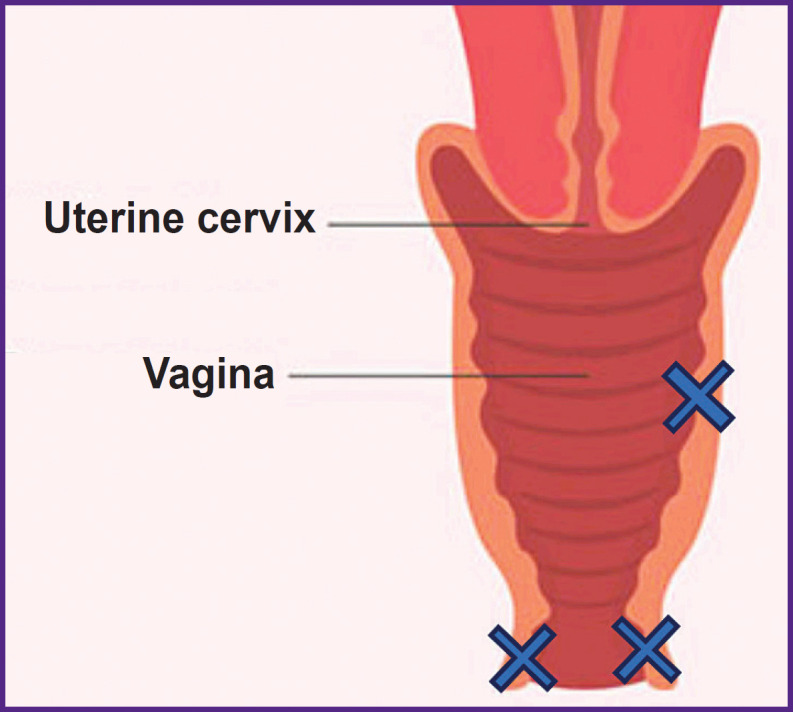
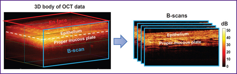
Materials and methods: The study involved 25 patients with clinical presentation of GUMS and 3 virtually healthy women (mean age - 56.7±1.4 years). On gynecological examination the patients underwent colpo- and vaginoscopy, their vaginal health index being calculated. OCT study was performed in three anatomical points of the upper vagina and the vaginal vestibule (these regions are rich in estrogen receptors and most frequently affected in GUSM). The biopsy was taken from the right point of the vaginal vestibule followed by a histological examination and PAS reaction to reveal glycogen. The epidermal thickness was quantitatively assessed by OCT and histological images. There were determined the signal levels from the epithelium and the connective tissue, the epithelial stromal stratification being performed.
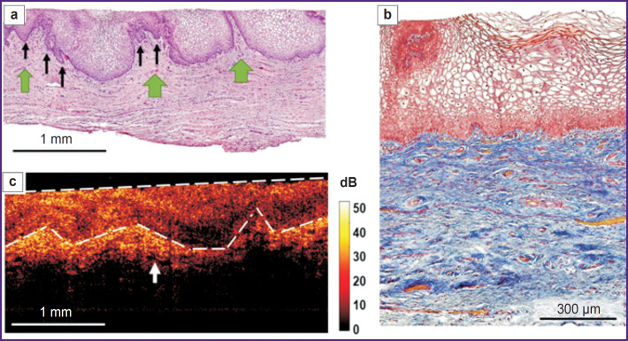
Results: Normal vaginal mucosa in OCT images had stratified structure including the epithelium 503 [467; 550] μm thick, with low intensity of OCT signal and the proper mucous plate with a high OCT signal. As a result of the difference in signal levels from the epithelium and the proper mucous plate, the contrast boundary formed between them. The signal level from the epithelium was 54.1 [51.5; 56.3] RU, and that from the connective tissue - 70.7 [65.9; 73.7] RU. The mucosa had folds, which in OCT images looked like a waveform boundary of the epithelium and the submucosa.Based on vaginal health index values and colpo- and vaginoscopy findings, GUSM patients were divided into 2 groups: patients with mild mucosal atrophy and those with severe mucosal atrophy. The first group of patients was observed to have the reduced epithelial thickness of up to 261 [244; 289] μm; the signal from the epithelium increased due to the decrease in glycogen content, and was 61.6 [55.0; 65.5] RU, and the connective tissue signal was 79.5 [77.2; 79.9] RU. Mucosal folds were not visible. Severe atrophy patients had a significant decrease in the epithelial thickness, up to 158 [143; 191] μm; the signal from the epithelium was 69.7 [67.1; 72.4] RU reducing the boundary contrast of the epithelium and the submucosal base (it can be explained by glycogen absence); the signal from the connective tissue was 90.32 [80.90; 101.60] RU. Mucosal folds were not visible. The stratification index showed no changes due to the fact that the signal intensity in vaginal atrophy increases synchronically from both: the epithelium and the proper plate (stroma). The epidermal thickness measured histologically showed a high coherence level with OCT measurements (r=0.93; p<0.0001).
Conclusion: The study determined OCT criteria of the age norm for vaginal mucosa changes and atrophy in mild and severe GUSM that will enable to facilitate the personalization of the therapy approaches and optimize the management of such patients.

 求助内容:
求助内容: 应助结果提醒方式:


