{"title":"Reproducibility of MRI-derived radiomic features in prostate cancer detection: a methodological approach.","authors":"Javad Zarei, Asma Soleimani, Marziyeh Tahmasbi, Mohsen Sarkarian, Seyed Masoud Rezaeijo","doi":"10.5114/pjr/201467","DOIUrl":null,"url":null,"abstract":"<p><strong>Purpose: </strong>We aim to evaluate the reproducibility of these features and apply machine learning algorithms to predict cancer diagnosis.</p><p><strong>Material and methods: </strong>We analyzed magnetic resonance (MR) images from a cohort of 82 individuals, split between 41 prostate cancer patients and 41 healthy controls. A total of 215 radiomic features were extracted from T2-weighted and ADC images using the Software Environment for Radiomic Analysis (SERA). Intraclass correlation coefficient (ICC) analysis was used to assess the reproducibility of features, and Pearson's correlation was applied to remove redundant features. After feature selection, seven dimensionality reduction techniques, including principal component analysis (PCA), kernel PCA, linear discriminant analysis, and locally linear embedding, were applied to preprocess the radiomic features. Ten machine learning algorithms, including support vector machines (SVM), random forests, neural networks, logistic regression, and ensemble methods such as CatBoost and AdaBoost, were utilized to classify cancerous versus non-cancerous tissues. Model performance was evaluated using accuracy and AUC-ROC metrics.</p><p><strong>Results: </strong>The results showed that features with high reproducibility (ICC > 0.75) contributed significantly to the performance of machine learning models. SVM, neural networks, and logistic regression achieved the highest accuracy (0.88-0.9) and AUC (up to 0.93) when using features from the good and excellent reproducibility categories. PCA emerged as the most effective dimensionality reduction method, preserving the discriminative power of reproducible features across all models.</p><p><strong>Conclusion: </strong>The results indicate that radiomic feature extraction from MR images, combined with dimensionality reduction and machine learning algorithms, provides a robust approach for prostate cancer diagnosis.</p>","PeriodicalId":94174,"journal":{"name":"Polish journal of radiology","volume":"90 ","pages":"e180-e188"},"PeriodicalIF":0.0000,"publicationDate":"2025-04-14","publicationTypes":"Journal Article","fieldsOfStudy":null,"isOpenAccess":false,"openAccessPdf":"https://www.ncbi.nlm.nih.gov/pmc/articles/PMC12099201/pdf/","citationCount":"0","resultStr":null,"platform":"Semanticscholar","paperid":null,"PeriodicalName":"Polish journal of radiology","FirstCategoryId":"1085","ListUrlMain":"https://doi.org/10.5114/pjr/201467","RegionNum":0,"RegionCategory":null,"ArticlePicture":[],"TitleCN":null,"AbstractTextCN":null,"PMCID":null,"EPubDate":"2025/1/1 0:00:00","PubModel":"eCollection","JCR":"","JCRName":"","Score":null,"Total":0}
引用次数: 0
Abstract
Purpose: We aim to evaluate the reproducibility of these features and apply machine learning algorithms to predict cancer diagnosis.
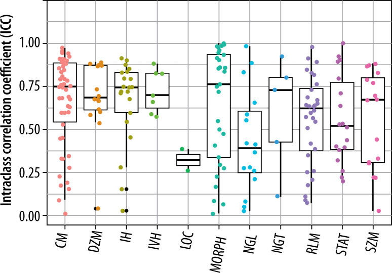
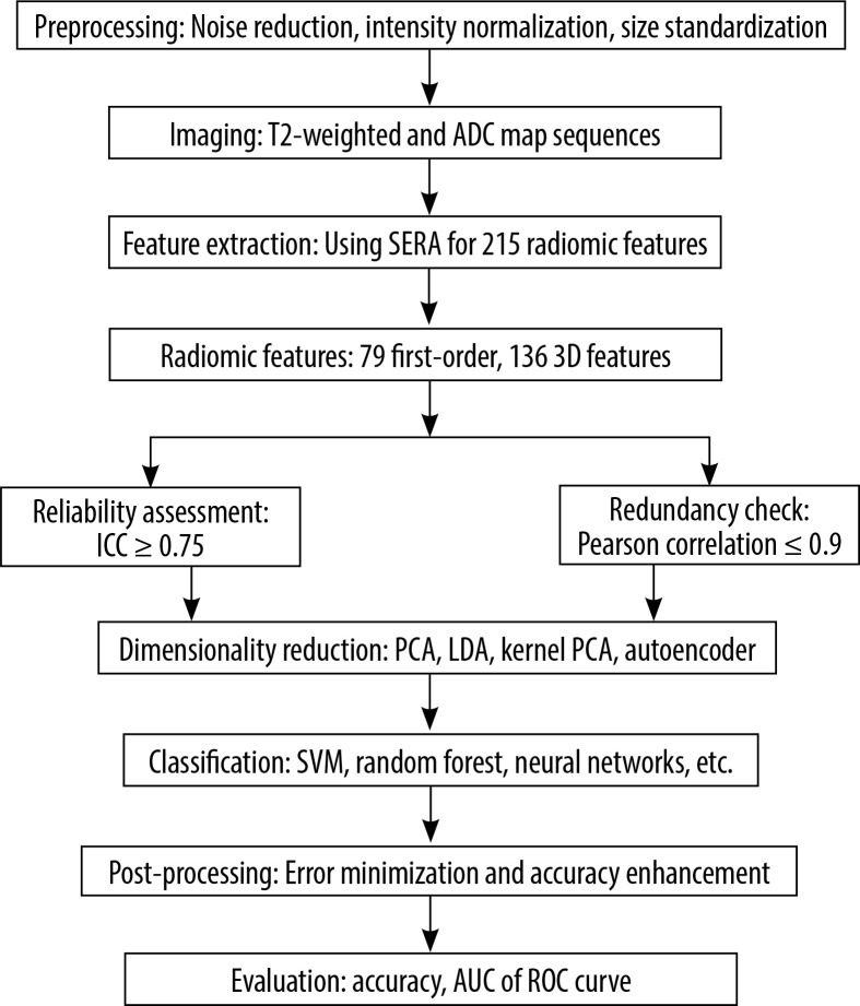
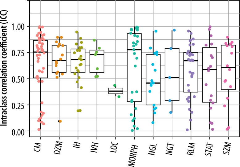
Material and methods: We analyzed magnetic resonance (MR) images from a cohort of 82 individuals, split between 41 prostate cancer patients and 41 healthy controls. A total of 215 radiomic features were extracted from T2-weighted and ADC images using the Software Environment for Radiomic Analysis (SERA). Intraclass correlation coefficient (ICC) analysis was used to assess the reproducibility of features, and Pearson's correlation was applied to remove redundant features. After feature selection, seven dimensionality reduction techniques, including principal component analysis (PCA), kernel PCA, linear discriminant analysis, and locally linear embedding, were applied to preprocess the radiomic features. Ten machine learning algorithms, including support vector machines (SVM), random forests, neural networks, logistic regression, and ensemble methods such as CatBoost and AdaBoost, were utilized to classify cancerous versus non-cancerous tissues. Model performance was evaluated using accuracy and AUC-ROC metrics.
Results: The results showed that features with high reproducibility (ICC > 0.75) contributed significantly to the performance of machine learning models. SVM, neural networks, and logistic regression achieved the highest accuracy (0.88-0.9) and AUC (up to 0.93) when using features from the good and excellent reproducibility categories. PCA emerged as the most effective dimensionality reduction method, preserving the discriminative power of reproducible features across all models.
Conclusion: The results indicate that radiomic feature extraction from MR images, combined with dimensionality reduction and machine learning algorithms, provides a robust approach for prostate cancer diagnosis.

 求助内容:
求助内容: 应助结果提醒方式:


