Burak Günay, Muzaffer Savaş Tepe, Halis Harun Öztürk, Atakan Küskün, Murat Gençbay
{"title":"Pericoronary fat attenuation in stenotic and vulnerable coronary artery plaques: Implications for coronary artery disease and associated conditions.","authors":"Burak Günay, Muzaffer Savaş Tepe, Halis Harun Öztürk, Atakan Küskün, Murat Gençbay","doi":"10.1177/20584601251342312","DOIUrl":null,"url":null,"abstract":"<p><strong>Background: </strong>Pericoronary adipose tissue density (PCAT) is a parameter that quantifies inflammation and atherosclerosis around the coronary arteries.</p><p><strong>Purpose: </strong>To investigate the correlation between PCAT and plaque features, stenosis degrees in coronary arteries (LAD, RCA, Cx) with stenotic vulnerable plaques.</p><p><strong>Material and methods: </strong>A Retrospective study including 103 patients (64M, 39F) who underwent coronary computed tomography was retrospectively examined at a single center. PCAT and high-risk plaques were measured independently and compared to stenosis and coronary artery type. Adipose tissue attenuation, ranging from -180 to -25 HU, was measured along the plaque's length and in a 0.5-1 mm region around the perilesional coronary arteries.</p><p><strong>Results: </strong>The PCAT values increases with the degree of stenosis in the LAD, Cx, and RCA (r = 0.9161, <i>p</i> < .001; r = 0.9717, <i>p</i> < .001; r = 0.9315, <i>p</i> < .001, respectively). PCAT values demonstrate a positive pattern when plaque length increases in all coronary arteries (r = -0.6316, <i>p</i> < .001; r = -0.8825, <i>p</i> < .001; r = -0.7529, <i>p</i> < .001; LAD, Cx, RCA). PCAT values differed significantly based on plaque type in all coronary arteries. Calcified plaques showed statistically significant differences compared to both soft and mixed plaques (<i>p</i> < .05). Patients with positive remodeling had PCAT values of -69.43 (±8.76) HU, while cases without positive remodeling had PCAT values of -84.54 (±7.65) HU, indicating a significant difference (<i>p</i> < .05).</p><p><strong>Conclusion: </strong>The combined evaluation of plaque features, stenosis degree, and PCAT provides a more accurate prediction of possible acute coronary syndrome cases than analyzing stenosis degree alone.</p>","PeriodicalId":72063,"journal":{"name":"Acta radiologica open","volume":"14 5","pages":"20584601251342312"},"PeriodicalIF":1.0000,"publicationDate":"2025-05-22","publicationTypes":"Journal Article","fieldsOfStudy":null,"isOpenAccess":false,"openAccessPdf":"https://www.ncbi.nlm.nih.gov/pmc/articles/PMC12099116/pdf/","citationCount":"0","resultStr":null,"platform":"Semanticscholar","paperid":null,"PeriodicalName":"Acta radiologica open","FirstCategoryId":"1085","ListUrlMain":"https://doi.org/10.1177/20584601251342312","RegionNum":0,"RegionCategory":null,"ArticlePicture":[],"TitleCN":null,"AbstractTextCN":null,"PMCID":null,"EPubDate":"2025/5/1 0:00:00","PubModel":"eCollection","JCR":"Q4","JCRName":"RADIOLOGY, NUCLEAR MEDICINE & MEDICAL IMAGING","Score":null,"Total":0}
引用次数: 0
Abstract
Background: Pericoronary adipose tissue density (PCAT) is a parameter that quantifies inflammation and atherosclerosis around the coronary arteries.
Purpose: To investigate the correlation between PCAT and plaque features, stenosis degrees in coronary arteries (LAD, RCA, Cx) with stenotic vulnerable plaques.
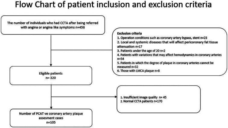
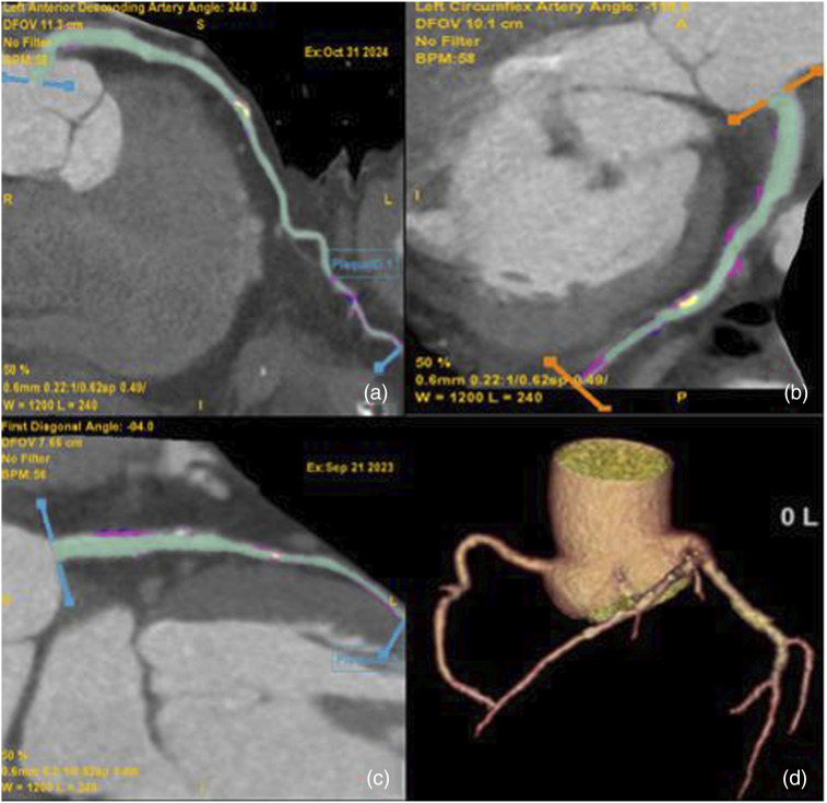
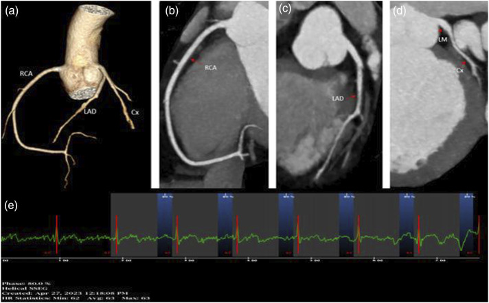
Material and methods: A Retrospective study including 103 patients (64M, 39F) who underwent coronary computed tomography was retrospectively examined at a single center. PCAT and high-risk plaques were measured independently and compared to stenosis and coronary artery type. Adipose tissue attenuation, ranging from -180 to -25 HU, was measured along the plaque's length and in a 0.5-1 mm region around the perilesional coronary arteries.
Results: The PCAT values increases with the degree of stenosis in the LAD, Cx, and RCA (r = 0.9161, p < .001; r = 0.9717, p < .001; r = 0.9315, p < .001, respectively). PCAT values demonstrate a positive pattern when plaque length increases in all coronary arteries (r = -0.6316, p < .001; r = -0.8825, p < .001; r = -0.7529, p < .001; LAD, Cx, RCA). PCAT values differed significantly based on plaque type in all coronary arteries. Calcified plaques showed statistically significant differences compared to both soft and mixed plaques (p < .05). Patients with positive remodeling had PCAT values of -69.43 (±8.76) HU, while cases without positive remodeling had PCAT values of -84.54 (±7.65) HU, indicating a significant difference (p < .05).
Conclusion: The combined evaluation of plaque features, stenosis degree, and PCAT provides a more accurate prediction of possible acute coronary syndrome cases than analyzing stenosis degree alone.

 求助内容:
求助内容: 应助结果提醒方式:


