Clinical and microbiological profile of patients with fungal keratitis demonstrating unusual yeast-like structures in potassium hydroxide with calcofluor white preparation of corneal scraping.
{"title":"Clinical and microbiological profile of patients with fungal keratitis demonstrating unusual yeast-like structures in potassium hydroxide with calcofluor white preparation of corneal scraping.","authors":"Sikha Misra, Savitri Sharma, Manas Ranjan Barik, Nisha Rani, Sujata Das, Srikant Kumar Sahu, Smruti Rekha Priyadarshini, Himansu Sekhar Behera","doi":"10.1186/s12348-025-00479-5","DOIUrl":null,"url":null,"abstract":"<p><strong>Background: </strong>To report a case series of microbial keratitis showing atypical yeast-like structures in direct microscopy which were culture-negative but subsequently identified as yeast cells by PCR and DNA sequencing.</p><p><strong>Main text: </strong>This is a retrospective, non-comparative case series of eight patients with infectious keratitis, where smear examination (potassium hydroxide + calcofluor white) showed spore like structures resembling yeast. There was no growth in any solid culture media. Routine PCR assay was performed using pan fungal primers followed by Sanger sequencing and nucleotide sequences were analysed using NCBI-BLAST software. Medical treatment in all patients were initiated based on clinical suspicion and presumptive microbiology report. Therapeutic penetrating keratoplasty was performed for patients not responding to medical antifungal therapy. Demographic, clinical data were collected for each patient from electronic medical records of the patients and outcome analysed. Amplification of fungal DNA was seen in the PCR assay of all samples. Nucleotide sequences of the amplicons obtained after Sanger sequencing and NCBI-BLAST analysis were found identical to Candida albicans (n = 7) and Citeromyces matriensis (n = 1). Patients were treated with antifungal drugs such as topical natamycin 5% or amphotericin B 0.15%. Ulcer resolved with scarring in 5 patients (62.5%), one patient had failed graft after therapeutic penetrating keratoplasty (12.5%), one (12.5%) eye became phthisical and one patient (12.5%) was lost to follow up.</p><p><strong>Conclusion: </strong>Atypical structures resembling yeast-like cells detected in direct smear examination in the absence of growth in culture media should raise the suspicion of a fungal etiology and warrant further investigations to establish the diagnosis.</p>","PeriodicalId":16600,"journal":{"name":"Journal of Ophthalmic Inflammation and Infection","volume":"15 1","pages":"45"},"PeriodicalIF":2.3000,"publicationDate":"2025-05-26","publicationTypes":"Journal Article","fieldsOfStudy":null,"isOpenAccess":false,"openAccessPdf":"https://www.ncbi.nlm.nih.gov/pmc/articles/PMC12106856/pdf/","citationCount":"0","resultStr":null,"platform":"Semanticscholar","paperid":null,"PeriodicalName":"Journal of Ophthalmic Inflammation and Infection","FirstCategoryId":"1085","ListUrlMain":"https://doi.org/10.1186/s12348-025-00479-5","RegionNum":0,"RegionCategory":null,"ArticlePicture":[],"TitleCN":null,"AbstractTextCN":null,"PMCID":null,"EPubDate":"","PubModel":"","JCR":"Q1","JCRName":"OPHTHALMOLOGY","Score":null,"Total":0}
引用次数: 0
Abstract
Background: To report a case series of microbial keratitis showing atypical yeast-like structures in direct microscopy which were culture-negative but subsequently identified as yeast cells by PCR and DNA sequencing.
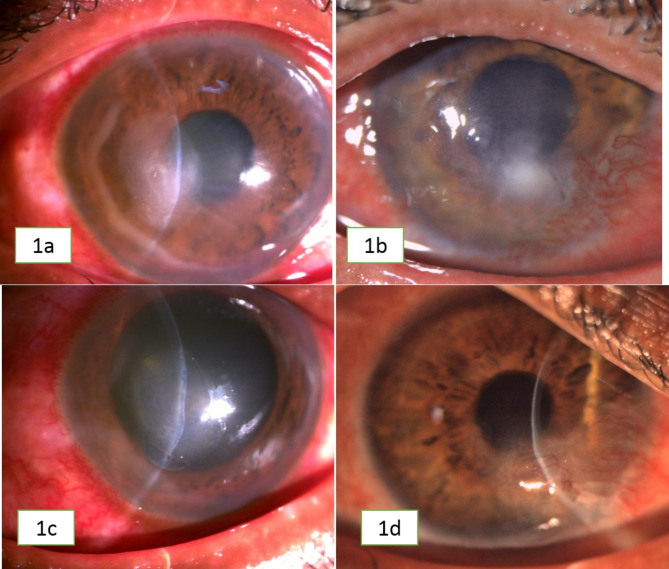
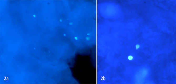
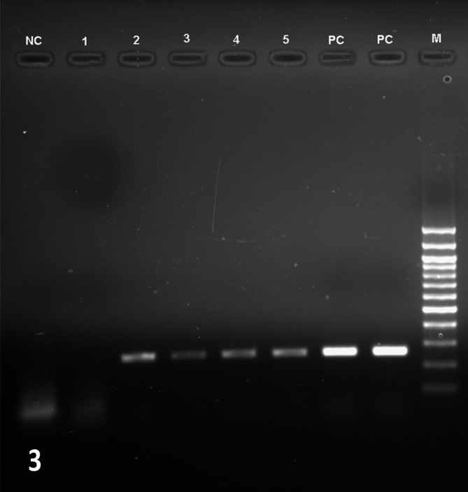
Main text: This is a retrospective, non-comparative case series of eight patients with infectious keratitis, where smear examination (potassium hydroxide + calcofluor white) showed spore like structures resembling yeast. There was no growth in any solid culture media. Routine PCR assay was performed using pan fungal primers followed by Sanger sequencing and nucleotide sequences were analysed using NCBI-BLAST software. Medical treatment in all patients were initiated based on clinical suspicion and presumptive microbiology report. Therapeutic penetrating keratoplasty was performed for patients not responding to medical antifungal therapy. Demographic, clinical data were collected for each patient from electronic medical records of the patients and outcome analysed. Amplification of fungal DNA was seen in the PCR assay of all samples. Nucleotide sequences of the amplicons obtained after Sanger sequencing and NCBI-BLAST analysis were found identical to Candida albicans (n = 7) and Citeromyces matriensis (n = 1). Patients were treated with antifungal drugs such as topical natamycin 5% or amphotericin B 0.15%. Ulcer resolved with scarring in 5 patients (62.5%), one patient had failed graft after therapeutic penetrating keratoplasty (12.5%), one (12.5%) eye became phthisical and one patient (12.5%) was lost to follow up.
Conclusion: Atypical structures resembling yeast-like cells detected in direct smear examination in the absence of growth in culture media should raise the suspicion of a fungal etiology and warrant further investigations to establish the diagnosis.
 求助内容:
求助内容: 应助结果提醒方式:


