Shalini Radhakrishnan, Nischitha N Suvarna, Saraswathy Sreeram, Srirama Bhat
{"title":"\"Colon\"ised by the unexpected: a case of extrauterine epithelioid trophoblastic tumour.","authors":"Shalini Radhakrishnan, Nischitha N Suvarna, Saraswathy Sreeram, Srirama Bhat","doi":"10.1186/s13000-025-01617-2","DOIUrl":null,"url":null,"abstract":"<p><strong>Introduction: </strong>Extrauterine epithelioid trophoblastic tumour is an exceedingly rare and aggressive form of gestational trophoblastic disease that arises outside the uterus and is characterised by the proliferation of intermediate trophoblastic cells. Unlike more common forms of gestational trophoblastic diseases, such as hydatidiform moles and choriocarcinoma, this entity presents unique diagnostic and therapeutic challenges due to its atypical location and clinical features. Thus far, no documented cases of this entity have been reported in the colon.</p><p><strong>Case presentation: </strong>We report the case of a 42-year-old woman who presented with complaints of lower abdominal pain and a palpable mass in the left iliac fossa, initially suspected to be an ectopic pregnancy. On radiological evaluation, a provisional diagnosis of gastrointestinal stromal tumour was made, following which the patient underwent a left colectomy with resection and anastomosis, and the excised specimen on comprehensive histopathological and immunohistochemical analysis was diagnosed as a case of extrauterine epithelioid trophoblastic tumour. However, the patient's condition deteriorated, and she succumbed to the disease one month after the diagnosis.</p><p><strong>Conclusion: </strong>The rarity of extrauterine trophoblastic tumours contributes to limited clinical experience and treatment protocols, resulting in poor prognoses. This case report highlights the importance of histopathological examination for a confirmatory diagnosis, ensuring timely identification and improving patient outcomes.</p>","PeriodicalId":11237,"journal":{"name":"Diagnostic Pathology","volume":"20 1","pages":"61"},"PeriodicalIF":2.3000,"publicationDate":"2025-05-24","publicationTypes":"Journal Article","fieldsOfStudy":null,"isOpenAccess":false,"openAccessPdf":"https://www.ncbi.nlm.nih.gov/pmc/articles/PMC12102896/pdf/","citationCount":"0","resultStr":null,"platform":"Semanticscholar","paperid":null,"PeriodicalName":"Diagnostic Pathology","FirstCategoryId":"3","ListUrlMain":"https://doi.org/10.1186/s13000-025-01617-2","RegionNum":3,"RegionCategory":"医学","ArticlePicture":[],"TitleCN":null,"AbstractTextCN":null,"PMCID":null,"EPubDate":"","PubModel":"","JCR":"Q2","JCRName":"PATHOLOGY","Score":null,"Total":0}
引用次数: 0
Abstract
Introduction: Extrauterine epithelioid trophoblastic tumour is an exceedingly rare and aggressive form of gestational trophoblastic disease that arises outside the uterus and is characterised by the proliferation of intermediate trophoblastic cells. Unlike more common forms of gestational trophoblastic diseases, such as hydatidiform moles and choriocarcinoma, this entity presents unique diagnostic and therapeutic challenges due to its atypical location and clinical features. Thus far, no documented cases of this entity have been reported in the colon.
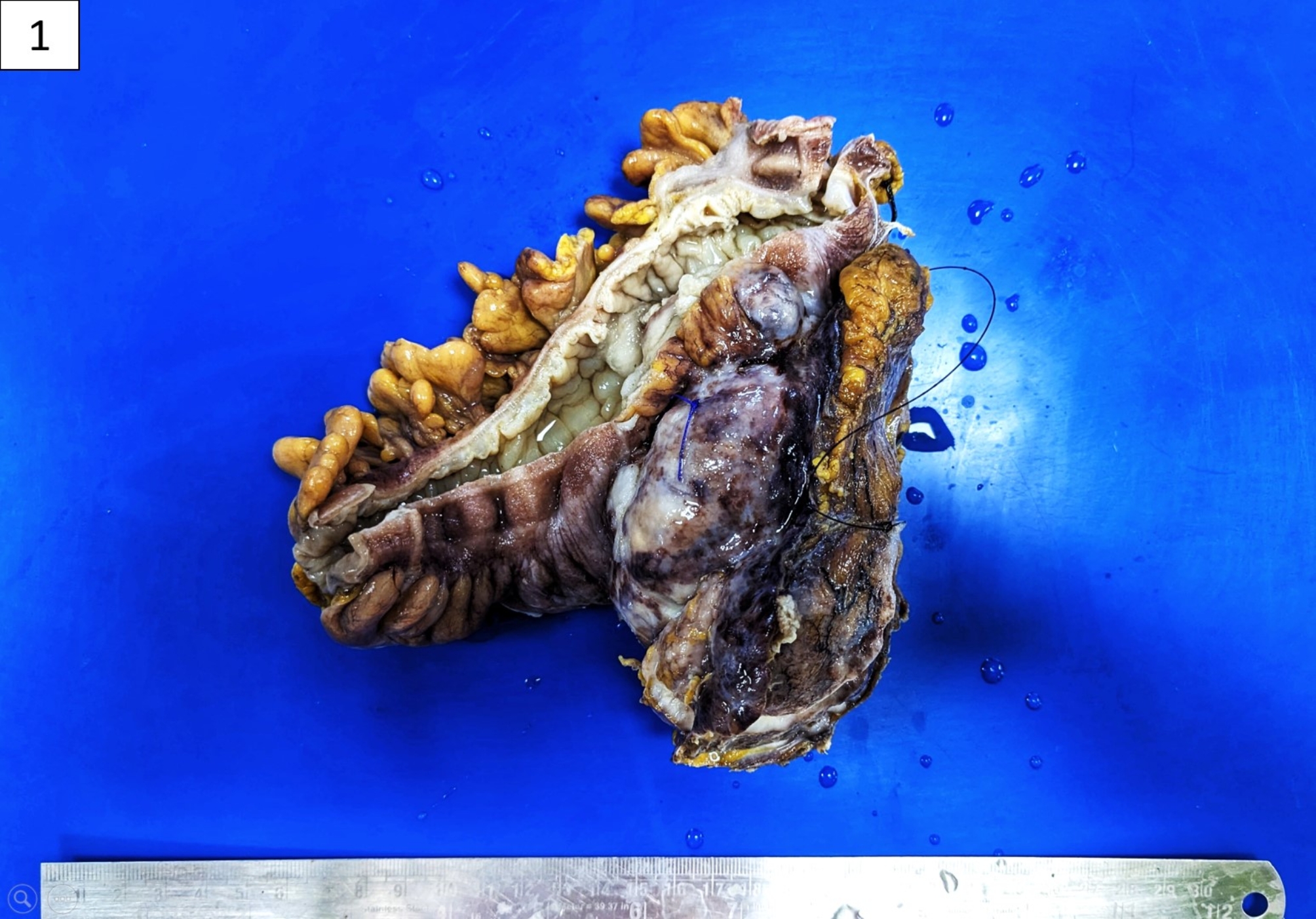
Case presentation: We report the case of a 42-year-old woman who presented with complaints of lower abdominal pain and a palpable mass in the left iliac fossa, initially suspected to be an ectopic pregnancy. On radiological evaluation, a provisional diagnosis of gastrointestinal stromal tumour was made, following which the patient underwent a left colectomy with resection and anastomosis, and the excised specimen on comprehensive histopathological and immunohistochemical analysis was diagnosed as a case of extrauterine epithelioid trophoblastic tumour. However, the patient's condition deteriorated, and she succumbed to the disease one month after the diagnosis.
Conclusion: The rarity of extrauterine trophoblastic tumours contributes to limited clinical experience and treatment protocols, resulting in poor prognoses. This case report highlights the importance of histopathological examination for a confirmatory diagnosis, ensuring timely identification and improving patient outcomes.
期刊介绍:
Diagnostic Pathology is an open access, peer-reviewed, online journal that considers research in surgical and clinical pathology, immunology, and biology, with a special focus on cutting-edge approaches in diagnostic pathology and tissue-based therapy. The journal covers all aspects of surgical pathology, including classic diagnostic pathology, prognosis-related diagnosis (tumor stages, prognosis markers, such as MIB-percentage, hormone receptors, etc.), and therapy-related findings. The journal also focuses on the technological aspects of pathology, including molecular biology techniques, morphometry aspects (stereology, DNA analysis, syntactic structure analysis), communication aspects (telecommunication, virtual microscopy, virtual pathology institutions, etc.), and electronic education and quality assurance (for example interactive publication, on-line references with automated updating, etc.).

 求助内容:
求助内容: 应助结果提醒方式:


