Florian Maria Bauer, Annette Sauerbeck, Wolfgang Hitzl, Nick Piravej, Josef Schmidbauer
{"title":"Automated Diabetic Retinopathy Screening in Out-patient Diabetes Care - Comparison of Two Artificial Intelligence Algorithms: RetCAD and OphtAI.","authors":"Florian Maria Bauer, Annette Sauerbeck, Wolfgang Hitzl, Nick Piravej, Josef Schmidbauer","doi":"10.1055/a-2620-1956","DOIUrl":null,"url":null,"abstract":"<p><strong>Objective: </strong>The artificial intelligence (AI) can be applied to screening for diabetic retinopathy (DR) from colour fundus photographs. The prerequisite for this is that the AI used can achieve a similar performance in the real world in different study conditions. The aim of this study is therefore to test and compare the latest version of the AI-based algorithms RetCAD and OphtAI for DR screening in a diabetes outpatient clinic.</p><p><strong>Methods: </strong>In the period from August 2023 to November 2023, 150 diabetics were recruited at the outpatient diabetes center of the University Hospital. For each study participant, images were taken with the handheld retinal camera Aurora (Optomed Plc, Oulu, Finland) in Miosis. The images were examined by the ophthalmologist and by the AI-based algorithms RetCAD version 2.2.0 (Thirona Retina, Nijmegen, Netherlands) and OphtAI version 2.3.4 (Groupe Evolucare Technologies, Le Pecq, France) for the presence of DR. The severity of DR was classified using the International Clinical Diabetic Retinopathy (ICDR) scale. Patients with no retinal changes or a mild DR were advised to have an ophthalmological check-up in one year. In the presence of a moderate, severe or proliferative DR, a referral to the treating ophthalmologist was made. For this reason, the severity levels of moderate, severe and proliferative DR have been summarised under the umbrella term of referable DR.</p><p><strong>Results: </strong>No DR was detected in 123 out of 143 (86.0%) diabetics and mild DR was detected in 10 (7.3%). All patients with moderate DR 7 (5.0%), severe 2 (1.5%) and proliferative DR 1 (0.7%) were grouped together as refererable DR and represented a proportion of 7.3%. The AI-based algorithm RetCAD version 2.2.0 achieved a sensitivity of 90% and a specificity of 100% for the detection of a referable DR compared to ophthalmological image assessment. RetCAD rated 98% of the images for image analysis as sufficient or better. In contrast, the second AI-based algorithm OphtAI version 2.3.4 achieved a sensitivity of 70% and a specificity of 100% for the detection of a referable DR. The OphtAI software was able to perform image analysis on all images.</p><p><strong>Conclusion: </strong>The results for the detection of a referable DR were consistent under study conditions and in clinical use for the AI-based algorithm RetCAD. The AI-based algorithm OphtAI, on the other hand, detected fewer patients with moderate DR, which was reflected in lower sensitivity. Both algorithms correctly assigned all patients with severe and proliferative DR. The AI-based algorithms RetCAD and OphtAI tested appear to be suitable for use in a diabetes outpatient clinic and primary care setting, respectively.</p>","PeriodicalId":17904,"journal":{"name":"Klinische Monatsblatter fur Augenheilkunde","volume":" ","pages":"912-919"},"PeriodicalIF":0.7000,"publicationDate":"2025-09-01","publicationTypes":"Journal Article","fieldsOfStudy":null,"isOpenAccess":false,"openAccessPdf":"https://www.ncbi.nlm.nih.gov/pmc/articles/PMC12445937/pdf/","citationCount":"0","resultStr":null,"platform":"Semanticscholar","paperid":null,"PeriodicalName":"Klinische Monatsblatter fur Augenheilkunde","FirstCategoryId":"3","ListUrlMain":"https://doi.org/10.1055/a-2620-1956","RegionNum":4,"RegionCategory":"医学","ArticlePicture":[],"TitleCN":null,"AbstractTextCN":null,"PMCID":null,"EPubDate":"2025/5/23 0:00:00","PubModel":"Epub","JCR":"Q4","JCRName":"OPHTHALMOLOGY","Score":null,"Total":0}
引用次数: 0
Abstract
Objective: The artificial intelligence (AI) can be applied to screening for diabetic retinopathy (DR) from colour fundus photographs. The prerequisite for this is that the AI used can achieve a similar performance in the real world in different study conditions. The aim of this study is therefore to test and compare the latest version of the AI-based algorithms RetCAD and OphtAI for DR screening in a diabetes outpatient clinic.
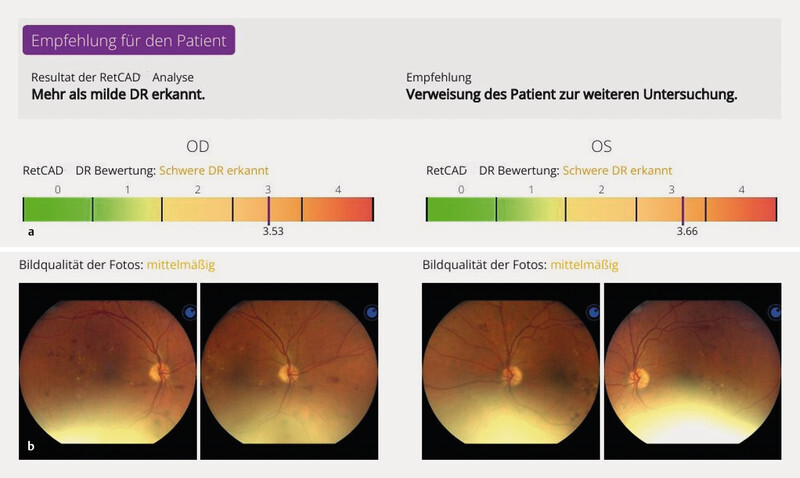
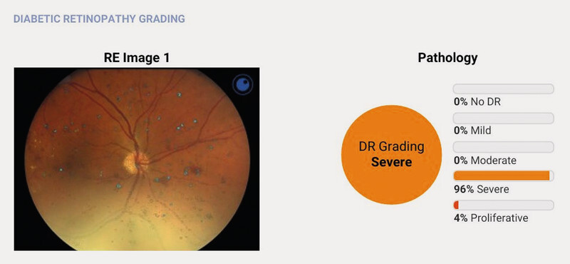
Methods: In the period from August 2023 to November 2023, 150 diabetics were recruited at the outpatient diabetes center of the University Hospital. For each study participant, images were taken with the handheld retinal camera Aurora (Optomed Plc, Oulu, Finland) in Miosis. The images were examined by the ophthalmologist and by the AI-based algorithms RetCAD version 2.2.0 (Thirona Retina, Nijmegen, Netherlands) and OphtAI version 2.3.4 (Groupe Evolucare Technologies, Le Pecq, France) for the presence of DR. The severity of DR was classified using the International Clinical Diabetic Retinopathy (ICDR) scale. Patients with no retinal changes or a mild DR were advised to have an ophthalmological check-up in one year. In the presence of a moderate, severe or proliferative DR, a referral to the treating ophthalmologist was made. For this reason, the severity levels of moderate, severe and proliferative DR have been summarised under the umbrella term of referable DR.
Results: No DR was detected in 123 out of 143 (86.0%) diabetics and mild DR was detected in 10 (7.3%). All patients with moderate DR 7 (5.0%), severe 2 (1.5%) and proliferative DR 1 (0.7%) were grouped together as refererable DR and represented a proportion of 7.3%. The AI-based algorithm RetCAD version 2.2.0 achieved a sensitivity of 90% and a specificity of 100% for the detection of a referable DR compared to ophthalmological image assessment. RetCAD rated 98% of the images for image analysis as sufficient or better. In contrast, the second AI-based algorithm OphtAI version 2.3.4 achieved a sensitivity of 70% and a specificity of 100% for the detection of a referable DR. The OphtAI software was able to perform image analysis on all images.
Conclusion: The results for the detection of a referable DR were consistent under study conditions and in clinical use for the AI-based algorithm RetCAD. The AI-based algorithm OphtAI, on the other hand, detected fewer patients with moderate DR, which was reflected in lower sensitivity. Both algorithms correctly assigned all patients with severe and proliferative DR. The AI-based algorithms RetCAD and OphtAI tested appear to be suitable for use in a diabetes outpatient clinic and primary care setting, respectively.
期刊介绍:
-Konzentriertes Fachwissen aus Klinik und Praxis:
Die entscheidenden Ergebnisse der internationalen Forschung - für Sie auf den Punkt gebracht und kritisch kommentiert,
Übersichtsarbeiten zu den maßgeblichen Themen der täglichen Praxis,
Top informiert - breite klinische Berichterstattung.
-CME-Punkte sammeln mit dem Refresher:
Effiziente, CME-zertifizierte Fortbildung, mit dem Refresher,
3 CME-Punkte pro Ausgabe - bis zu 36 CME-Punkte im Jahr!.
-Aktuelle Rubriken mit echtem Nutzwert:
Kurzreferate zu den wichtigsten Artikeln internationaler Zeitschriften,
Schwerpunktthema in jedem Heft: Ausführliche Übersichtsarbeiten zu den wichtigsten Themen der Ophthalmologie – so behalten Sie das gesamte Fach im Blick!,
Originalien mit den neuesten Entwicklungen,
Übersichten zu den relevanten Themen.

 求助内容:
求助内容: 应助结果提醒方式:


