Deep learning-based automatic differentiation of acute angle closure with or without zonulopathy using ultrasound biomicroscopy: a comparison of diagnostic performance with ophthalmologists.
{"title":"Deep learning-based automatic differentiation of acute angle closure with or without zonulopathy using ultrasound biomicroscopy: a comparison of diagnostic performance with ophthalmologists.","authors":"Lu Cheng, Jiaming Hong, Hailiu Chen, Yunlan Ling, Shufen Lin, Jing Huang, Ethan Wu, Yangyunhui Li, Haotian Lin, Shaopeng Liu, Jingjing Huang","doi":"10.1136/bmjophth-2024-002114","DOIUrl":null,"url":null,"abstract":"<p><strong>Objective: </strong>This study aims to develop ultrasound biomicroscopy (UBM)-based artificial intelligence (AI) models for preoperative differentiation of acute angle closure (AAC) with or without zonulopathy and to compare their comprehensive diagnostic performance against ophthalmologists as a cross-sectional study.</p><p><strong>Methods and analysis: </strong>Three AI models were developed to differentiate AAC with or without zonular laxity or lens subluxation using UBM images and ocular parameters. Their diagnostic performances were analysed, with the best-performing model then compared with two diagnostic methods used by ophthalmologists (logistic regression and UBM image analysis). Additionally, a robustness validation dataset, including images from UBM and anterior segment optical coherence tomography (AS-OCT), was used to validate the robustness of the best-performing AI model.</p><p><strong>Results: </strong>A total of 537 eyes were included in this study. The best-performing AI model was image-based and achieved a macro-area under the curve (AUC) of 0.9046 with a diagnostic processing time of 0.03 s per image in differentiating AAC with or without zonulopathy. The manually calculated multinomial logistic regression model achieved a macro-AUC of 0.9373, requiring 1200.00 s per analysis. UBM image analysis achieved a mean accuracy and processing time of 64.17% and 20.13 s, respectively, per image. Robustness validation of the image-based AI model showed an accuracy of 66.67% and 61.11% for UBM and AS-OCT images.</p><p><strong>Conclusions: </strong>AI models and ophthalmologists effectively differentiated AAC with or without zonulopathy. However, when evaluated in terms of both accuracy and efficiency, the AI model showed superior comprehensive diagnostic performance, demonstrating high clinical applicability for preoperative diagnosis.</p>","PeriodicalId":9286,"journal":{"name":"BMJ Open Ophthalmology","volume":"10 1","pages":""},"PeriodicalIF":2.2000,"publicationDate":"2025-05-22","publicationTypes":"Journal Article","fieldsOfStudy":null,"isOpenAccess":false,"openAccessPdf":"https://www.ncbi.nlm.nih.gov/pmc/articles/PMC12104886/pdf/","citationCount":"0","resultStr":null,"platform":"Semanticscholar","paperid":null,"PeriodicalName":"BMJ Open Ophthalmology","FirstCategoryId":"1085","ListUrlMain":"https://doi.org/10.1136/bmjophth-2024-002114","RegionNum":0,"RegionCategory":null,"ArticlePicture":[],"TitleCN":null,"AbstractTextCN":null,"PMCID":null,"EPubDate":"","PubModel":"","JCR":"Q2","JCRName":"OPHTHALMOLOGY","Score":null,"Total":0}
引用次数: 0
Abstract
Objective: This study aims to develop ultrasound biomicroscopy (UBM)-based artificial intelligence (AI) models for preoperative differentiation of acute angle closure (AAC) with or without zonulopathy and to compare their comprehensive diagnostic performance against ophthalmologists as a cross-sectional study.
Methods and analysis: Three AI models were developed to differentiate AAC with or without zonular laxity or lens subluxation using UBM images and ocular parameters. Their diagnostic performances were analysed, with the best-performing model then compared with two diagnostic methods used by ophthalmologists (logistic regression and UBM image analysis). Additionally, a robustness validation dataset, including images from UBM and anterior segment optical coherence tomography (AS-OCT), was used to validate the robustness of the best-performing AI model.
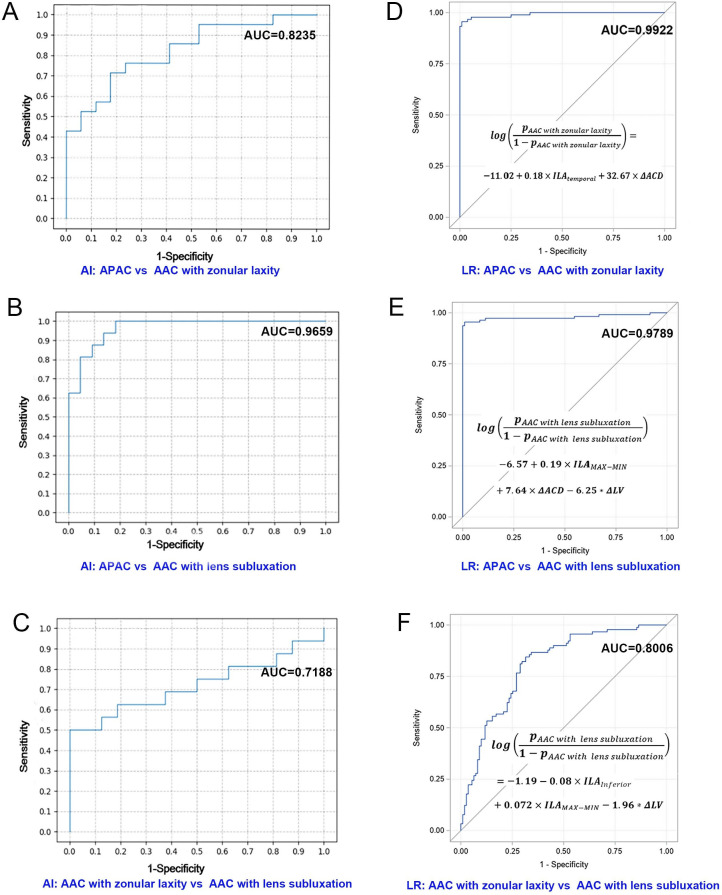
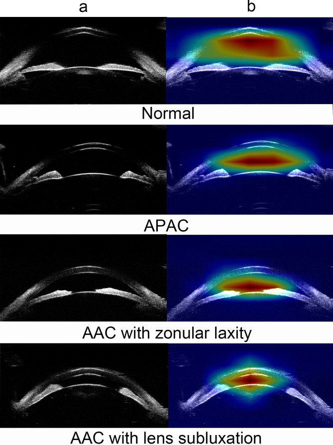
Results: A total of 537 eyes were included in this study. The best-performing AI model was image-based and achieved a macro-area under the curve (AUC) of 0.9046 with a diagnostic processing time of 0.03 s per image in differentiating AAC with or without zonulopathy. The manually calculated multinomial logistic regression model achieved a macro-AUC of 0.9373, requiring 1200.00 s per analysis. UBM image analysis achieved a mean accuracy and processing time of 64.17% and 20.13 s, respectively, per image. Robustness validation of the image-based AI model showed an accuracy of 66.67% and 61.11% for UBM and AS-OCT images.
Conclusions: AI models and ophthalmologists effectively differentiated AAC with or without zonulopathy. However, when evaluated in terms of both accuracy and efficiency, the AI model showed superior comprehensive diagnostic performance, demonstrating high clinical applicability for preoperative diagnosis.

 求助内容:
求助内容: 应助结果提醒方式:


