Mihaly Enyedi, Georgian-Theodor Badea, Radu-Tudor Ion, Daniela Elena Gheoca Mutu, Stefan Oprea, Zoran Florin Filipoiu
{"title":"The dissection-based identification of the preaortic sympathetic plexus formation, anatomical relations, and clinical applications.","authors":"Mihaly Enyedi, Georgian-Theodor Badea, Radu-Tudor Ion, Daniela Elena Gheoca Mutu, Stefan Oprea, Zoran Florin Filipoiu","doi":"10.25122/jml-2025-0069","DOIUrl":null,"url":null,"abstract":"<p><p>The abdominal sympathetic nervous system provides sympathetic innervation to the abdominal organs and gonads. This system is part of an extensive neural network that extends from the base of the skull to the pelvis. The preaortic (or prevertebral) plexus is a key component of the abdominal sympathetic system and is represented by a variable nervous network located anterior to the abdominal aorta. The aim of our study was to identify all these sympathetic structures and describe the formation and relationships of the preaortic plexus. We examined five cadavers (aged 66-71) with no medical or surgical history, preserved in 9% formalin at the Anatomy Department from Carol Davila University. Regional dissections were performed in successive planes, highlighting the major abdominal plexuses, the lumbar splanchnic nerves, and the associated network of neural connections that contribute to the preaortic plexus. The plexus is formed by efferent fibers from the celiac and aortico-renal ganglia, as well as from the three lumbar splanchnic nerves. The lumbar splanchnic nerves originate in the paravertebral sympathetic chains. We identified all these sympathetic structures and described the formation and anatomical relationships of the plexus. The nerve fibers of various origins form a longitudinally oriented network located anterolateral to the abdominal aorta. The lower part of this network continues into the superior hypogastric plexus. This neural network is delicate, complex, and variable, making it challenging to identify anatomically and surgically. Situated deeply in the retroperitoneal space, it is prone to accidental injuries during surgery in this compartment.</p>","PeriodicalId":16386,"journal":{"name":"Journal of Medicine and Life","volume":"18 4","pages":"351-356"},"PeriodicalIF":0.0000,"publicationDate":"2025-04-01","publicationTypes":"Journal Article","fieldsOfStudy":null,"isOpenAccess":false,"openAccessPdf":"https://www.ncbi.nlm.nih.gov/pmc/articles/PMC12094316/pdf/","citationCount":"0","resultStr":null,"platform":"Semanticscholar","paperid":null,"PeriodicalName":"Journal of Medicine and Life","FirstCategoryId":"1085","ListUrlMain":"https://doi.org/10.25122/jml-2025-0069","RegionNum":0,"RegionCategory":null,"ArticlePicture":[],"TitleCN":null,"AbstractTextCN":null,"PMCID":null,"EPubDate":"","PubModel":"","JCR":"Q3","JCRName":"Medicine","Score":null,"Total":0}
引用次数: 0
Abstract
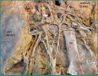
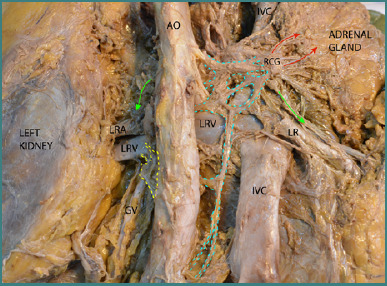
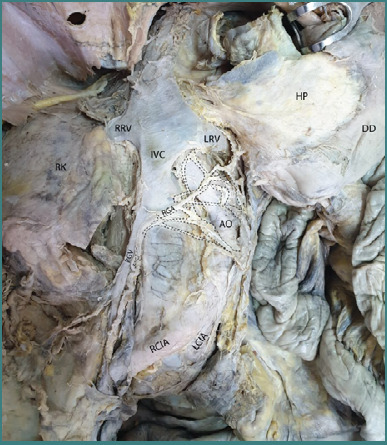
The abdominal sympathetic nervous system provides sympathetic innervation to the abdominal organs and gonads. This system is part of an extensive neural network that extends from the base of the skull to the pelvis. The preaortic (or prevertebral) plexus is a key component of the abdominal sympathetic system and is represented by a variable nervous network located anterior to the abdominal aorta. The aim of our study was to identify all these sympathetic structures and describe the formation and relationships of the preaortic plexus. We examined five cadavers (aged 66-71) with no medical or surgical history, preserved in 9% formalin at the Anatomy Department from Carol Davila University. Regional dissections were performed in successive planes, highlighting the major abdominal plexuses, the lumbar splanchnic nerves, and the associated network of neural connections that contribute to the preaortic plexus. The plexus is formed by efferent fibers from the celiac and aortico-renal ganglia, as well as from the three lumbar splanchnic nerves. The lumbar splanchnic nerves originate in the paravertebral sympathetic chains. We identified all these sympathetic structures and described the formation and anatomical relationships of the plexus. The nerve fibers of various origins form a longitudinally oriented network located anterolateral to the abdominal aorta. The lower part of this network continues into the superior hypogastric plexus. This neural network is delicate, complex, and variable, making it challenging to identify anatomically and surgically. Situated deeply in the retroperitoneal space, it is prone to accidental injuries during surgery in this compartment.
期刊介绍:
The Journal of Medicine and Life publishes peer-reviewed articles from various fields of medicine and life sciences, including original research, systematic reviews, special reports, case presentations, major medical breakthroughs and letters to the editor. The Journal focuses on current matters that lie at the intersection of biomedical science and clinical practice and strives to present this information to inform health care delivery and improve patient outcomes. Papers addressing topics such as neuroprotection, neurorehabilitation, neuroplasticity, and neuroregeneration are particularly encouraged, as part of the Journal''s continuous interest in neuroscience research. The Editorial Board of the Journal of Medicine and Life is open to consider manuscripts from all levels of research and areas of biological sciences, including fundamental, experimental or clinical research and matters of public health. As part of our pledge to promote an educational and community-building environment, our issues feature sections designated to informing our readers regarding exciting international congresses, teaching courses and relevant institutional-level events.

 求助内容:
求助内容: 应助结果提醒方式:


