{"title":"Neuronal activity triggers secretory autophagy to mediate the extracellular release of SNCA/α-synuclein.","authors":"Yoshitsugu Nakamura, Shigeki Arawaka","doi":"10.1080/27694127.2024.2410683","DOIUrl":null,"url":null,"abstract":"<p><p>Autophagy has two distinct pathways, degradation and secretion. Autophagic degradation plays a pivotal role in cellular homeostasis by the formation of a double-membrane autophagosome in concert with numerous ATG (autophagy-related) proteins. However, the mechanism that mediates autophagic secretion is not fully understood. To explore how autophagic secretion is physiologically triggered and regulated in neurons, we investigated whether neuronal activity affected autophagic secretion by analyzing SNCA secretion in mouse primary cortical neurons and SH-SY5Y cells. In primary neurons, rapamycin promoted SNCA secretion, while the effect was canceled in primary neurons of <i>Becn1</i> <sup>+/-</sup>deficient mice. Stimulating neuronal activity by glutamate promoted SNCA secretion, autophagic flux, and colocalization of SNCA with LC3 (microtubule-associated proteins 1 light chain 3). These effects were inhibited by the intracellular Ca<sup>2+</sup> chelator BAPTA-AM. Additionally, glutamate-induced SNCA secretion was suppressed by <i>Atg5</i> or <i>Rab8a</i> knockdown in SH-SY5Y cells, and mainly occurred in the fashion associated with extracellular vesicles in primary neurons. These results suggest that neuronal activity triggers autophagic secretion for releasing SNCA via an increase in intracellular Ca<sup>2+</sup> concentration.</p>","PeriodicalId":72341,"journal":{"name":"Autophagy reports","volume":"3 1","pages":"2410683"},"PeriodicalIF":0.0000,"publicationDate":"2024-10-07","publicationTypes":"Journal Article","fieldsOfStudy":null,"isOpenAccess":false,"openAccessPdf":"https://www.ncbi.nlm.nih.gov/pmc/articles/PMC11864697/pdf/","citationCount":"0","resultStr":null,"platform":"Semanticscholar","paperid":null,"PeriodicalName":"Autophagy reports","FirstCategoryId":"1085","ListUrlMain":"https://doi.org/10.1080/27694127.2024.2410683","RegionNum":0,"RegionCategory":null,"ArticlePicture":[],"TitleCN":null,"AbstractTextCN":null,"PMCID":null,"EPubDate":"2024/1/1 0:00:00","PubModel":"eCollection","JCR":"","JCRName":"","Score":null,"Total":0}
引用次数: 0
Abstract
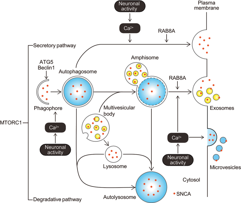
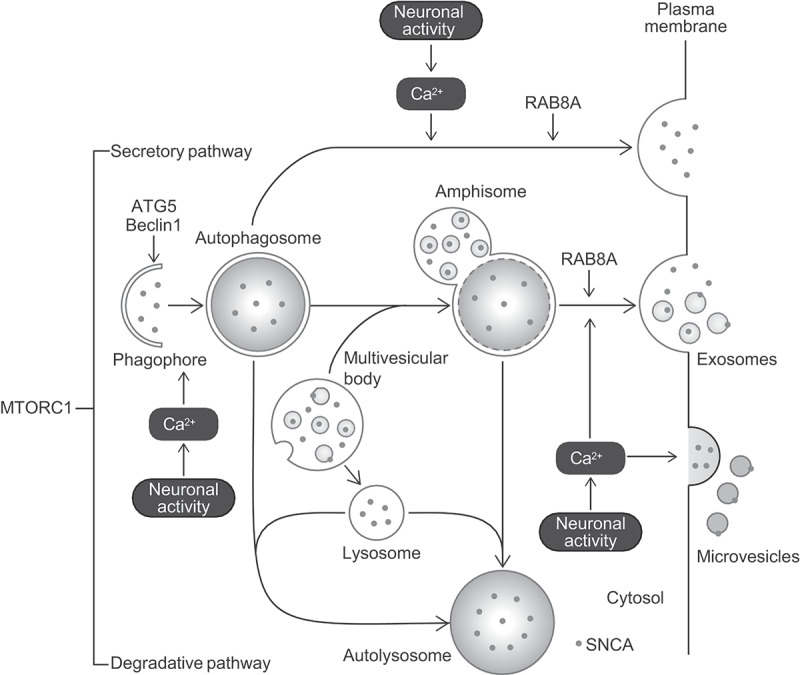
Autophagy has two distinct pathways, degradation and secretion. Autophagic degradation plays a pivotal role in cellular homeostasis by the formation of a double-membrane autophagosome in concert with numerous ATG (autophagy-related) proteins. However, the mechanism that mediates autophagic secretion is not fully understood. To explore how autophagic secretion is physiologically triggered and regulated in neurons, we investigated whether neuronal activity affected autophagic secretion by analyzing SNCA secretion in mouse primary cortical neurons and SH-SY5Y cells. In primary neurons, rapamycin promoted SNCA secretion, while the effect was canceled in primary neurons of Becn1+/-deficient mice. Stimulating neuronal activity by glutamate promoted SNCA secretion, autophagic flux, and colocalization of SNCA with LC3 (microtubule-associated proteins 1 light chain 3). These effects were inhibited by the intracellular Ca2+ chelator BAPTA-AM. Additionally, glutamate-induced SNCA secretion was suppressed by Atg5 or Rab8a knockdown in SH-SY5Y cells, and mainly occurred in the fashion associated with extracellular vesicles in primary neurons. These results suggest that neuronal activity triggers autophagic secretion for releasing SNCA via an increase in intracellular Ca2+ concentration.

 求助内容:
求助内容: 应助结果提醒方式:


