The impact of dilated cardiomyopathy in relation to coronary artery dimensions and left ventricle myocardial mass in a model with excluded coronary atherosclerosis.
Jarosław Skowroński, Emilia Szudejko, Adam Banasiak, Kacper Milczanowski, Paweł Jelski, Ilona Michałowska, Cezary Kępka, Mariusz Kruk, Adam Witkowski, Jerzy Pręgowski
{"title":"The impact of dilated cardiomyopathy in relation to coronary artery dimensions and left ventricle myocardial mass in a model with excluded coronary atherosclerosis.","authors":"Jarosław Skowroński, Emilia Szudejko, Adam Banasiak, Kacper Milczanowski, Paweł Jelski, Ilona Michałowska, Cezary Kępka, Mariusz Kruk, Adam Witkowski, Jerzy Pręgowski","doi":"10.5603/cj.104850","DOIUrl":null,"url":null,"abstract":"<p><strong>Background: </strong>The diagnosis of dilated cardiomyopathy (DCM) requires exclusion of obstructive coronary artery disease (CAD). However, co-occurrence of DCM and non-obstructive coronary atherosclerotic plaque is frequent. Our objective was to evaluate the coronary artery dimensions and their relation to the left ventricle mass in DCM patients with excluded coronary atherosclerosis.</p><p><strong>Methods: </strong>Out of 426 patients with DCM who underwent computed coronary tomography angiography (CCTA), we identified 34 without signs of coronary atherosclerosis and compared them with 193 consecutive patients without DCM and atherosclerosis in CCTA. They were matched one to three by sex, coronary dominance pattern, and body-surface area (BSA). Left ventricle myocardial mass (LVMM) and proximal and middle coronary artery segment dimensions were evaluated with the use of dedicated, commercially available software.</p><p><strong>Results: </strong>Overall, coronary segment dimensions were not different between groups except for the medial left anterior descending segment and obtuse marginal, which were wider in the DCM group, while the proximal right coronary artery was larger in the non-DCM group. Total coronary artery volume (CAV) and LVMM/CAV ratio were greater in the DCM group (2879 [2535-3508] mm³ vs. 2521 [2120-3115] mm³, p = 0.03) and (0.062 [0.054-0.074] g/mm³ vs. 0.049 [0.039-0.058] mm³, p = 0.0002), respectively. Also, the LVMM/coronary artery ostial area (COA) ratio was larger in patients with DCM (5.4±1.3 g/mm² vs. 3.7 ± 1.1 g/mm², p < 0.0001). The independent positive predictors of a larger LVMM/CAV ratio were DCM and BSA, while age was a negative predictor. LVMM/COA ratio positive predictors were DCM and male sex.</p><p><strong>Conclusions: </strong>Patients with DCM have altered relationships between LVMM, CAV, and COA.</p>","PeriodicalId":93923,"journal":{"name":"Cardiology journal","volume":" ","pages":"369-379"},"PeriodicalIF":0.0000,"publicationDate":"2025-01-01","publicationTypes":"Journal Article","fieldsOfStudy":null,"isOpenAccess":false,"openAccessPdf":"https://www.ncbi.nlm.nih.gov/pmc/articles/PMC12410942/pdf/","citationCount":"0","resultStr":null,"platform":"Semanticscholar","paperid":null,"PeriodicalName":"Cardiology journal","FirstCategoryId":"1085","ListUrlMain":"https://doi.org/10.5603/cj.104850","RegionNum":0,"RegionCategory":null,"ArticlePicture":[],"TitleCN":null,"AbstractTextCN":null,"PMCID":null,"EPubDate":"2025/5/20 0:00:00","PubModel":"Epub","JCR":"","JCRName":"","Score":null,"Total":0}
引用次数: 0
Abstract
Background: The diagnosis of dilated cardiomyopathy (DCM) requires exclusion of obstructive coronary artery disease (CAD). However, co-occurrence of DCM and non-obstructive coronary atherosclerotic plaque is frequent. Our objective was to evaluate the coronary artery dimensions and their relation to the left ventricle mass in DCM patients with excluded coronary atherosclerosis.
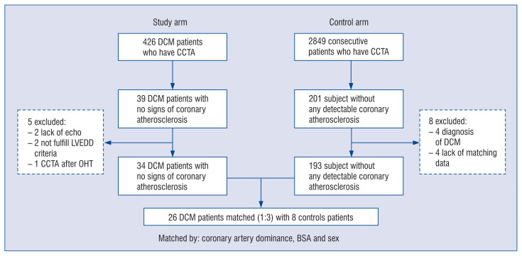
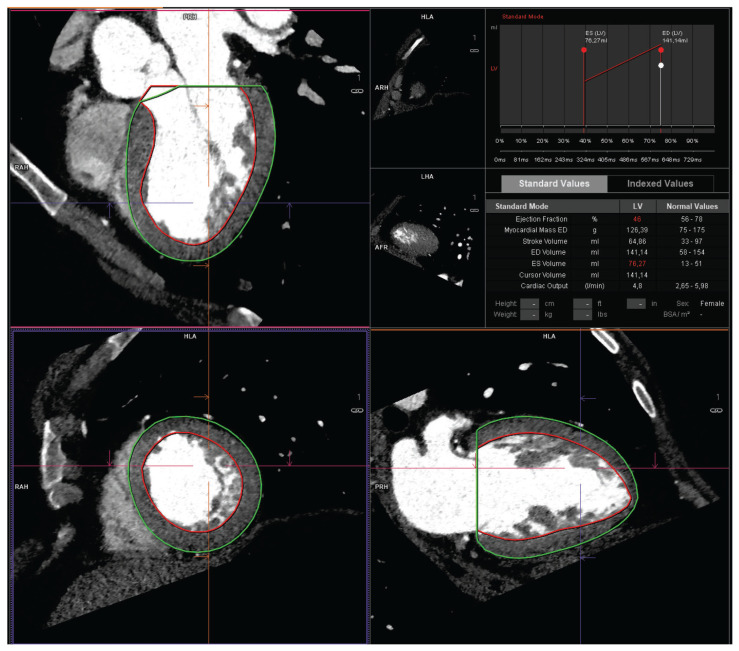
Methods: Out of 426 patients with DCM who underwent computed coronary tomography angiography (CCTA), we identified 34 without signs of coronary atherosclerosis and compared them with 193 consecutive patients without DCM and atherosclerosis in CCTA. They were matched one to three by sex, coronary dominance pattern, and body-surface area (BSA). Left ventricle myocardial mass (LVMM) and proximal and middle coronary artery segment dimensions were evaluated with the use of dedicated, commercially available software.
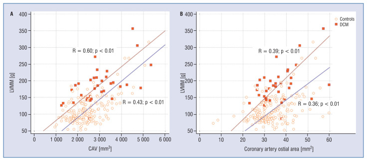
Results: Overall, coronary segment dimensions were not different between groups except for the medial left anterior descending segment and obtuse marginal, which were wider in the DCM group, while the proximal right coronary artery was larger in the non-DCM group. Total coronary artery volume (CAV) and LVMM/CAV ratio were greater in the DCM group (2879 [2535-3508] mm³ vs. 2521 [2120-3115] mm³, p = 0.03) and (0.062 [0.054-0.074] g/mm³ vs. 0.049 [0.039-0.058] mm³, p = 0.0002), respectively. Also, the LVMM/coronary artery ostial area (COA) ratio was larger in patients with DCM (5.4±1.3 g/mm² vs. 3.7 ± 1.1 g/mm², p < 0.0001). The independent positive predictors of a larger LVMM/CAV ratio were DCM and BSA, while age was a negative predictor. LVMM/COA ratio positive predictors were DCM and male sex.
Conclusions: Patients with DCM have altered relationships between LVMM, CAV, and COA.

 求助内容:
求助内容: 应助结果提醒方式:


