Nikolaus Wachtel, Luisa Weber, Nicholas Moellhoff, Constanze Kuhlmann, Riccardo E Giunta, Paolo Alberton, Denis Ehrl, Severin Wiggenhauser
{"title":"Platelet-Rich Fibrin Mediates Beneficial Effects on Adipose-Derived Stem Cells via Increased Levels of Key Cytokines.","authors":"Nikolaus Wachtel, Luisa Weber, Nicholas Moellhoff, Constanze Kuhlmann, Riccardo E Giunta, Paolo Alberton, Denis Ehrl, Severin Wiggenhauser","doi":"10.1111/wrr.70040","DOIUrl":null,"url":null,"abstract":"<p><p>Recent studies showcased the regenerative potential of Platelet-Rich Fibrin (PRF) combined with Adipose-Derived Stem Cells (ASC). PRF enhances cellular proliferation through sustained growth factor secretion which are continuously released to surrounding cells. However, its regulatory mechanisms remain unclear. ASC were isolated from liposuction and abdominoplasty samples of healthy donors, characterised via flow-cytometry and cultured for 7 days. Four cell culture conditions were tested: (1) 10% PRF extract (PRFe), (2) 10% Platelet-Low Plasma (PLP), (3) 10% Foetal Bovine Serum (FBS) and (4) basal medium as control. Cell viability and proliferation were assessed using AlamarBlue and PicoGreen assays, as well as live-dead staining. Enzyme-Linked Immunosorbent Assays quantified growth factor concentrations, while multiplex qPCR and immunocytochemical staining analysed gene and protein expression on days 1 and 7. PRFe-supplemented cultures showed the highest viability and proliferation, significantly surpassing other groups at day 7 (p < 0.05). Supernatant analysis revealed significantly elevated TGF-β1 and PDGF-AA/BB levels in PRFe cultures at day 7 (p of at least < 0.05). Multiplex qPCR indicated increased expression of proliferation and pluripotency markers (NANOG, JUN, SOX2, RPS6KA4; p < 0.05) and fibrillar collagen (COL1A; p < 0.05) in the PRFe group. These findings demonstrate that PRFe significantly enhances ASC proliferation and regenerative potential. Elevated levels of TGF-1, PDGF-AA/BB and to a lesser extend VEGF in PRFe cultures suggest that its benefits in regenerative medicine may be linked to these cytokines' upregulation. These results underscore PRFe's potential as a key supplement for optimising ASC-based therapies in tissue regeneration.</p>","PeriodicalId":23864,"journal":{"name":"Wound Repair and Regeneration","volume":"33 3","pages":"e70040"},"PeriodicalIF":3.4000,"publicationDate":"2025-05-01","publicationTypes":"Journal Article","fieldsOfStudy":null,"isOpenAccess":false,"openAccessPdf":"https://www.ncbi.nlm.nih.gov/pmc/articles/PMC12093288/pdf/","citationCount":"0","resultStr":null,"platform":"Semanticscholar","paperid":null,"PeriodicalName":"Wound Repair and Regeneration","FirstCategoryId":"3","ListUrlMain":"https://doi.org/10.1111/wrr.70040","RegionNum":3,"RegionCategory":"医学","ArticlePicture":[],"TitleCN":null,"AbstractTextCN":null,"PMCID":null,"EPubDate":"","PubModel":"","JCR":"Q2","JCRName":"CELL BIOLOGY","Score":null,"Total":0}
引用次数: 0
Abstract
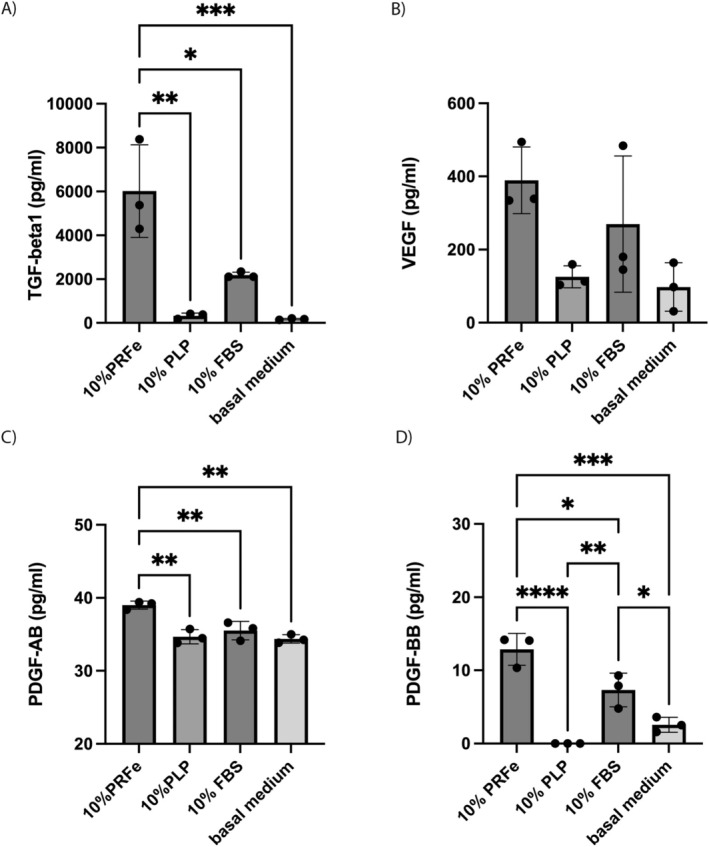
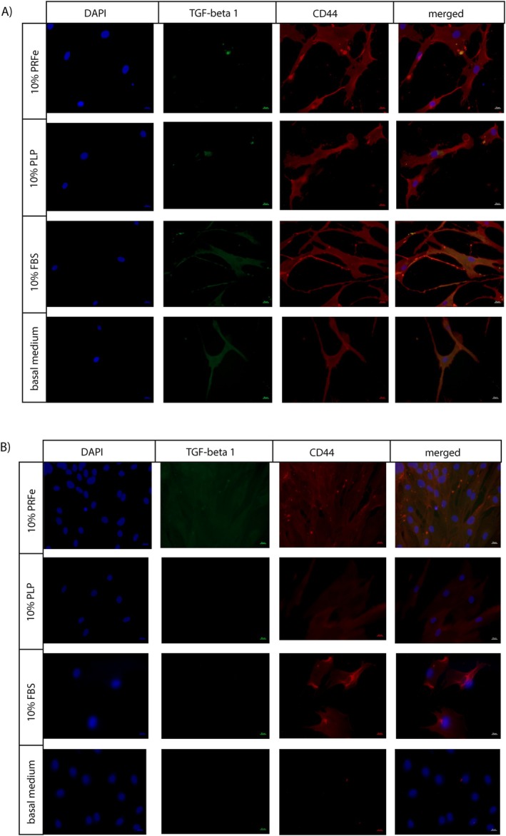
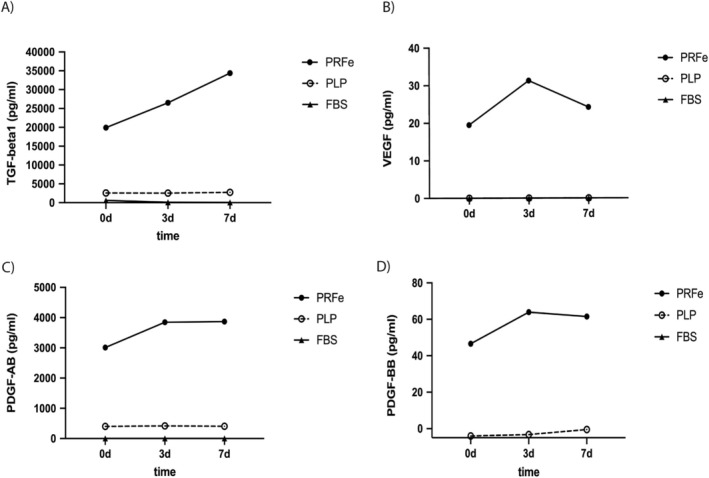
Recent studies showcased the regenerative potential of Platelet-Rich Fibrin (PRF) combined with Adipose-Derived Stem Cells (ASC). PRF enhances cellular proliferation through sustained growth factor secretion which are continuously released to surrounding cells. However, its regulatory mechanisms remain unclear. ASC were isolated from liposuction and abdominoplasty samples of healthy donors, characterised via flow-cytometry and cultured for 7 days. Four cell culture conditions were tested: (1) 10% PRF extract (PRFe), (2) 10% Platelet-Low Plasma (PLP), (3) 10% Foetal Bovine Serum (FBS) and (4) basal medium as control. Cell viability and proliferation were assessed using AlamarBlue and PicoGreen assays, as well as live-dead staining. Enzyme-Linked Immunosorbent Assays quantified growth factor concentrations, while multiplex qPCR and immunocytochemical staining analysed gene and protein expression on days 1 and 7. PRFe-supplemented cultures showed the highest viability and proliferation, significantly surpassing other groups at day 7 (p < 0.05). Supernatant analysis revealed significantly elevated TGF-β1 and PDGF-AA/BB levels in PRFe cultures at day 7 (p of at least < 0.05). Multiplex qPCR indicated increased expression of proliferation and pluripotency markers (NANOG, JUN, SOX2, RPS6KA4; p < 0.05) and fibrillar collagen (COL1A; p < 0.05) in the PRFe group. These findings demonstrate that PRFe significantly enhances ASC proliferation and regenerative potential. Elevated levels of TGF-1, PDGF-AA/BB and to a lesser extend VEGF in PRFe cultures suggest that its benefits in regenerative medicine may be linked to these cytokines' upregulation. These results underscore PRFe's potential as a key supplement for optimising ASC-based therapies in tissue regeneration.
期刊介绍:
Wound Repair and Regeneration provides extensive international coverage of cellular and molecular biology, connective tissue, and biological mediator studies in the field of tissue repair and regeneration and serves a diverse audience of surgeons, plastic surgeons, dermatologists, biochemists, cell biologists, and others.
Wound Repair and Regeneration is the official journal of The Wound Healing Society, The European Tissue Repair Society, The Japanese Society for Wound Healing, and The Australian Wound Management Association.
 求助内容:
求助内容: 应助结果提醒方式:


