Honglan Mi, Philipp Boehm-Sturm, Akvile Haeckel, Ying Li, Susanne Mueller, Fei Ni, Harald Kratz, Marco Foddis, Jing Xie, Eyk Schellenberger
{"title":"High-resolution quantitative mapping of extracellular pH by ratiometric MRI with iron chelates in a tumor mouse model.","authors":"Honglan Mi, Philipp Boehm-Sturm, Akvile Haeckel, Ying Li, Susanne Mueller, Fei Ni, Harald Kratz, Marco Foddis, Jing Xie, Eyk Schellenberger","doi":"10.1007/s11547-025-02020-z","DOIUrl":null,"url":null,"abstract":"<p><strong>Purpose: </strong>The aim of this study was to generate quantitative extracellular pH maps of tumors using a combination of a pH-sensitive iron chelate-based contrast agent (IBCA) and a pH-insensitive IBCA for concentration measurement, which we termed ratiometric pH magnetic resonance imaging (RpH-MRI).</p><p><strong>Methods: </strong>The pH-sensitive IBCA of ethylenediamine-trans-cyclohexane diamine tetraacetic acid (Fe-en-tCDTA) was synthesized, along with the pH-insensitive IBCAs of trans-cyclohexane diamine tetraacetic acid (Fe-tCDTA) and diethylenetriamine-N,N,N',N″,N″-pentaacetic acid (Fe-DTPA). The pH-dependent T1 contrast effects of these chelates were compared in water and serum phantoms at 0.94 T, 3 T and 7 T. For in vivo pH mapping of tumors at 7 T, 4T1 breast cancer cells were inoculated subcutaneously into the flanks of the BALB/c mice. RpH-MRI was performed with two sequential intravenous applications: first a pH-insensitive IBCA, followed by the pH-sensitive IBCA at the same dose (0.25 or 0.5 mmol/kg) with an interval of either 30 or 60 min. Quantitative pH maps were generated by calculating T1, S<sub>0</sub>, and relative maximum enhancement maps of the two injections, together with pH-dependent T1-relaxivity parameters derived from in vitro measurements of the pH-sensitive IBCA and pH-insensitive control IBCA.</p><p><strong>Results: </strong>The T1 relaxivity (r1) of Fe-en-tCDTA was highly pH dependent, being approximately 2.7 times higher at pH 5.5 than at neutral pH, whereas Fe-DTPA and Fe-tCDTA showed stable r1 values between pH 5.5-7.4. In vivo, the time to maximum signal intensity (TMI) of the tumors of Fe-DTPA as control was comparable to that of Fe-en-tCDTA (2.57 ± 1.34 min vs. 2.683 ± 0.89 min, p = 0.7596, paired t test, 4 mice, 7 tumors) as well as for Fe-tCDTA as control versus Fe-en-tCDTA (3.30 ± 1.17 min vs. 3.627 ± 1.12 min, p = 0.2101, paired t test, 7 mice, 13 tumors), suggesting similar pharmacokinetics. The concentration distribution at TMI of the control chelates was assumed to be the same as that of the second injected Fe-en-tCDTA. The dynamic contrast enhanced MRI curve of the first injection of Fe-DTPA returned to baseline after 20-30 min, whereas Fe-tCDTA took 30-60 min to reach baseline. Calculated core and rim pH values were 6.512 ± 0.182 and 6.742 ± 0.121, respectively (p < 0.0001, paired t test, 11 mice, 20 tumors) with core areas showing lower chelate concentrations but higher T1 relaxivity; the mean tumor-wide pH value was 6.632 ± 0.140.</p><p><strong>Conclusion: </strong>Our results demonstrate the potential of high-resolution RpH-MRI based on pH-sensitive and pH-insensitive IBCAs for mapping tumor extracellular pH and concentration distribution.</p>","PeriodicalId":20817,"journal":{"name":"Radiologia Medica","volume":" ","pages":"1231-1242"},"PeriodicalIF":4.8000,"publicationDate":"2025-08-01","publicationTypes":"Journal Article","fieldsOfStudy":null,"isOpenAccess":false,"openAccessPdf":"https://www.ncbi.nlm.nih.gov/pmc/articles/PMC12367910/pdf/","citationCount":"0","resultStr":null,"platform":"Semanticscholar","paperid":null,"PeriodicalName":"Radiologia Medica","FirstCategoryId":"3","ListUrlMain":"https://doi.org/10.1007/s11547-025-02020-z","RegionNum":1,"RegionCategory":"医学","ArticlePicture":[],"TitleCN":null,"AbstractTextCN":null,"PMCID":null,"EPubDate":"2025/5/20 0:00:00","PubModel":"Epub","JCR":"Q1","JCRName":"RADIOLOGY, NUCLEAR MEDICINE & MEDICAL IMAGING","Score":null,"Total":0}
引用次数: 0
Abstract
Purpose: The aim of this study was to generate quantitative extracellular pH maps of tumors using a combination of a pH-sensitive iron chelate-based contrast agent (IBCA) and a pH-insensitive IBCA for concentration measurement, which we termed ratiometric pH magnetic resonance imaging (RpH-MRI).
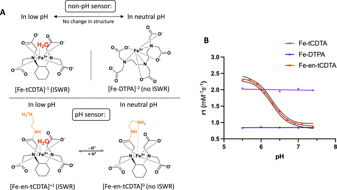
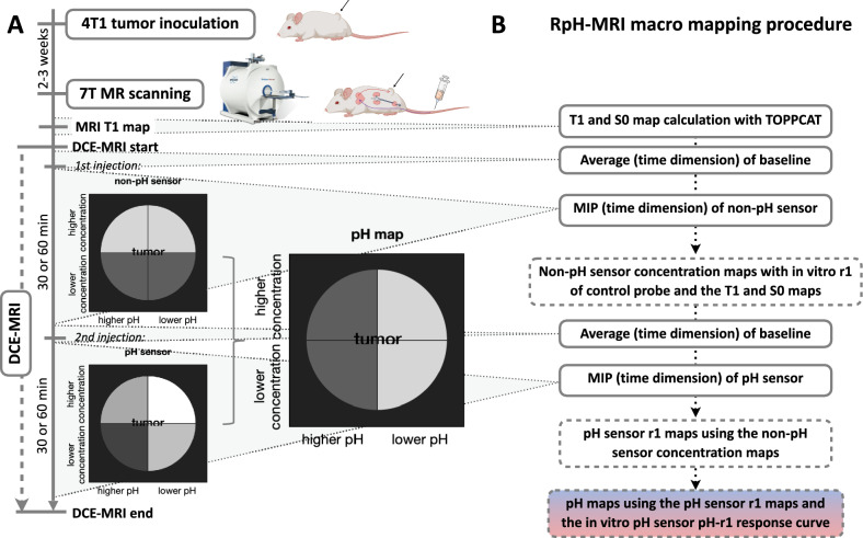
Methods: The pH-sensitive IBCA of ethylenediamine-trans-cyclohexane diamine tetraacetic acid (Fe-en-tCDTA) was synthesized, along with the pH-insensitive IBCAs of trans-cyclohexane diamine tetraacetic acid (Fe-tCDTA) and diethylenetriamine-N,N,N',N″,N″-pentaacetic acid (Fe-DTPA). The pH-dependent T1 contrast effects of these chelates were compared in water and serum phantoms at 0.94 T, 3 T and 7 T. For in vivo pH mapping of tumors at 7 T, 4T1 breast cancer cells were inoculated subcutaneously into the flanks of the BALB/c mice. RpH-MRI was performed with two sequential intravenous applications: first a pH-insensitive IBCA, followed by the pH-sensitive IBCA at the same dose (0.25 or 0.5 mmol/kg) with an interval of either 30 or 60 min. Quantitative pH maps were generated by calculating T1, S0, and relative maximum enhancement maps of the two injections, together with pH-dependent T1-relaxivity parameters derived from in vitro measurements of the pH-sensitive IBCA and pH-insensitive control IBCA.
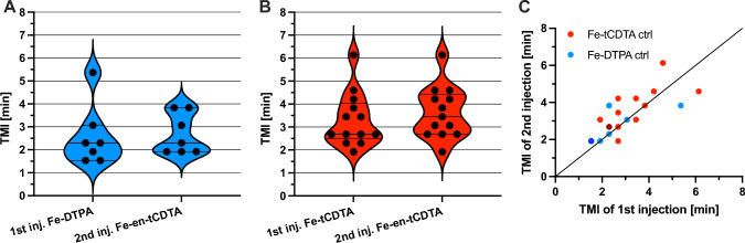
Results: The T1 relaxivity (r1) of Fe-en-tCDTA was highly pH dependent, being approximately 2.7 times higher at pH 5.5 than at neutral pH, whereas Fe-DTPA and Fe-tCDTA showed stable r1 values between pH 5.5-7.4. In vivo, the time to maximum signal intensity (TMI) of the tumors of Fe-DTPA as control was comparable to that of Fe-en-tCDTA (2.57 ± 1.34 min vs. 2.683 ± 0.89 min, p = 0.7596, paired t test, 4 mice, 7 tumors) as well as for Fe-tCDTA as control versus Fe-en-tCDTA (3.30 ± 1.17 min vs. 3.627 ± 1.12 min, p = 0.2101, paired t test, 7 mice, 13 tumors), suggesting similar pharmacokinetics. The concentration distribution at TMI of the control chelates was assumed to be the same as that of the second injected Fe-en-tCDTA. The dynamic contrast enhanced MRI curve of the first injection of Fe-DTPA returned to baseline after 20-30 min, whereas Fe-tCDTA took 30-60 min to reach baseline. Calculated core and rim pH values were 6.512 ± 0.182 and 6.742 ± 0.121, respectively (p < 0.0001, paired t test, 11 mice, 20 tumors) with core areas showing lower chelate concentrations but higher T1 relaxivity; the mean tumor-wide pH value was 6.632 ± 0.140.
Conclusion: Our results demonstrate the potential of high-resolution RpH-MRI based on pH-sensitive and pH-insensitive IBCAs for mapping tumor extracellular pH and concentration distribution.
目的:本研究的目的是使用pH敏感铁螯合造影剂(IBCA)和pH不敏感IBCA的组合来生成肿瘤的定量细胞外pH图,我们称之为比率pH磁共振成像(RpH-MRI)。方法:合成对ph敏感的乙二胺-反式环己烷二胺四乙酸(Fe-en-tCDTA)、对ph不敏感的乙二胺-四乙酸(Fe-tCDTA)和二乙三胺-N,N,N',N″,N″-五乙酸(Fe-DTPA)的IBCA。在0.94 T、3 T和7 T的水和血清幻影中比较这些螯合剂的pH依赖性T1对比效应。为了在7 T时肿瘤的体内pH定位,将4T1乳腺癌细胞皮下接种到BALB/c小鼠的侧壁。通过连续两次静脉注射进行RpH-MRI:首先是pH不敏感的IBCA,然后是相同剂量(0.25或0.5 mmol/kg)的pH敏感的IBCA,间隔30或60分钟。通过计算两次注射的T1、S0和相对最大增强图,以及从pH敏感IBCA和pH不敏感对照IBCA的体外测量得出的pH依赖的T1松弛度参数,生成定量pH图。结果:Fe-en-tCDTA的T1弛豫度(r1)高度依赖于pH值,pH为5.5时约为中性pH时的2.7倍,而Fe-DTPA和Fe-tCDTA的T1弛豫度(r1)在pH为5.5-7.4之间保持稳定。在体内,Fe-DTPA组与Fe-en-tCDTA组达到肿瘤最大信号强度(TMI)的时间相当(2.57±1.34 min vs. 2.683±0.89 min, p = 0.7596,配对t检验,4只小鼠,7个肿瘤),Fe-tCDTA组与Fe-en-tCDTA组相比(3.30±1.17 min vs. 3.627±1.12 min, p = 0.2101,配对t检验,7只小鼠,13个肿瘤),表明相似的药代动力学。假设对照螯合物在TMI处的浓度分布与第二次注射的Fe-en-tCDTA相同。首次注射Fe-DTPA的动态增强MRI曲线在20- 30min后恢复到基线,而Fe-tCDTA需要30- 60min才能达到基线。计算的核心和边缘pH值分别为6.512±0.182和6.742±0.121 (p)。结论:基于pH敏感和pH不敏感的IBCAs,高分辨率RpH-MRI在绘制肿瘤细胞外pH和浓度分布方面具有潜力。
期刊介绍:
Felice Perussia founded La radiologia medica in 1914. It is a peer-reviewed journal and serves as the official journal of the Italian Society of Medical and Interventional Radiology (SIRM). The primary purpose of the journal is to disseminate information related to Radiology, especially advancements in diagnostic imaging and related disciplines. La radiologia medica welcomes original research on both fundamental and clinical aspects of modern radiology, with a particular focus on diagnostic and interventional imaging techniques. It also covers topics such as radiotherapy, nuclear medicine, radiobiology, health physics, and artificial intelligence in the context of clinical implications. The journal includes various types of contributions such as original articles, review articles, editorials, short reports, and letters to the editor. With an esteemed Editorial Board and a selection of insightful reports, the journal is an indispensable resource for radiologists and professionals in related fields. Ultimately, La radiologia medica aims to serve as a platform for international collaboration and knowledge sharing within the radiological community.

 求助内容:
求助内容: 应助结果提醒方式:


