Jeong Hun Park, Nettie E Brown, Sarah Jo Tucker, Johnna S Temenoff, Mark El-Deiry, Hyun-Ji Park, Andrew T Tkaczuk, Scott J Hollister
{"title":"Feasibility Assessment of 3D Printing-Based Tubular Tissue Flap in a Porcine Model for Long Segmental Tracheal Reconstruction.","authors":"Jeong Hun Park, Nettie E Brown, Sarah Jo Tucker, Johnna S Temenoff, Mark El-Deiry, Hyun-Ji Park, Andrew T Tkaczuk, Scott J Hollister","doi":"10.1007/s13770-025-00718-9","DOIUrl":null,"url":null,"abstract":"<p><strong>Background: </strong>Despite advances in tissue engineering, current clinical reconstructive options for long segment tracheal defects are limited. In this study, a 3D printing based tubular tissue flap strategy was developed for long segment tracheal reconstruction.</p><p><strong>Method: </strong>A stent-patterned airway scaffold with sufficient radial rigidity and longitudinal bending flexibility was designed and its mechanical behavior was analyzed using finite element analysis (FEA). The stent-patterned airway scaffolds with a removable central core to preserve an internal lumen were created by selective laser sintering (SLS) based 3D printing. The stent-patterned airway scaffold with the central core, filled with poly (ethylene glycol) diacrylate-dithiothreitol (PEGDA-DTT) hydrogel containing erythropoietin (EPO) to enhance vascularization, was then implanted into the latissimus dorsi muscle of a Yucatan minipig.</p><p><strong>Results: </strong>A tubular tissue flap, with controlled luminal layer thickness was successfully created by removing the central core from the retrieved tissue flap containing the airway scaffold after 45 days of implantation in the Yucatan minipig model.</p><p><strong>Conclusion: </strong>The current work validated the potential of the tubular tissue flap based on the 3D printing as a clinically viable tissue engineering strategy for long segment tracheal reconstruction.</p>","PeriodicalId":23126,"journal":{"name":"Tissue engineering and regenerative medicine","volume":" ","pages":"469-479"},"PeriodicalIF":4.1000,"publicationDate":"2025-06-01","publicationTypes":"Journal Article","fieldsOfStudy":null,"isOpenAccess":false,"openAccessPdf":"https://www.ncbi.nlm.nih.gov/pmc/articles/PMC12122989/pdf/","citationCount":"0","resultStr":null,"platform":"Semanticscholar","paperid":null,"PeriodicalName":"Tissue engineering and regenerative medicine","FirstCategoryId":"5","ListUrlMain":"https://doi.org/10.1007/s13770-025-00718-9","RegionNum":4,"RegionCategory":"医学","ArticlePicture":[],"TitleCN":null,"AbstractTextCN":null,"PMCID":null,"EPubDate":"2025/5/21 0:00:00","PubModel":"Epub","JCR":"Q2","JCRName":"CELL & TISSUE ENGINEERING","Score":null,"Total":0}
引用次数: 0
Abstract
Background: Despite advances in tissue engineering, current clinical reconstructive options for long segment tracheal defects are limited. In this study, a 3D printing based tubular tissue flap strategy was developed for long segment tracheal reconstruction.
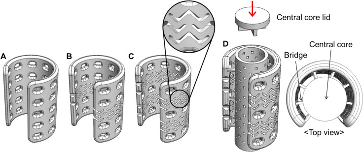
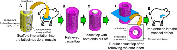
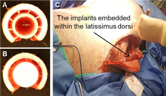
Method: A stent-patterned airway scaffold with sufficient radial rigidity and longitudinal bending flexibility was designed and its mechanical behavior was analyzed using finite element analysis (FEA). The stent-patterned airway scaffolds with a removable central core to preserve an internal lumen were created by selective laser sintering (SLS) based 3D printing. The stent-patterned airway scaffold with the central core, filled with poly (ethylene glycol) diacrylate-dithiothreitol (PEGDA-DTT) hydrogel containing erythropoietin (EPO) to enhance vascularization, was then implanted into the latissimus dorsi muscle of a Yucatan minipig.
Results: A tubular tissue flap, with controlled luminal layer thickness was successfully created by removing the central core from the retrieved tissue flap containing the airway scaffold after 45 days of implantation in the Yucatan minipig model.
Conclusion: The current work validated the potential of the tubular tissue flap based on the 3D printing as a clinically viable tissue engineering strategy for long segment tracheal reconstruction.
期刊介绍:
Tissue Engineering and Regenerative Medicine (Tissue Eng Regen Med, TERM), the official journal of the Korean Tissue Engineering and Regenerative Medicine Society, is a publication dedicated to providing research- based solutions to issues related to human diseases. This journal publishes articles that report substantial information and original findings on tissue engineering, medical biomaterials, cells therapy, stem cell biology and regenerative medicine.
 求助内容:
求助内容: 应助结果提醒方式:


