Philipp Becker, Andreas Pabst, Diana Heimes, Nadine Wiesmann-Imilowski, Sven Schumann, Peer W Kämmerer
{"title":"Biomechanical and cellular assessment of novel partially demineralized allogeneic bone plates: an ex-vivo and in-vitro study.","authors":"Philipp Becker, Andreas Pabst, Diana Heimes, Nadine Wiesmann-Imilowski, Sven Schumann, Peer W Kämmerer","doi":"10.1186/s40729-025-00625-7","DOIUrl":null,"url":null,"abstract":"<p><strong>Purpose: </strong>This study aimed to compare commercial allogeneic cortical bone plates (cCP) with innovative, differently demineralized CP (dCP) in biomechanics and human osteoblast (HOB) viability ex-vivo and in-vitro.</p><p><strong>Methods: </strong>Breaking strength (BS; in N) and flexibility (F; in mm) of cCP and dCP were assessed and compared using four groups ((1) non-hydrated, (2) hydrated for 10, (3) 30, and (4) 60 min in saline), respectively. Cell viability of HOB was evaluated by resazurin reduction on non-hydrated cCP and dCP after 3, 7, and 10 days. Scanning electron microscopy (SEM) visualized CP breaking edges, internal structures, HOB cell morphology, and growth patterns.</p><p><strong>Results: </strong>BS of hydrated dCP (d10: 15.45 ± 7.01 N, d30: 19.40 ± 3.78 N, d60: 20.31 ± 4.90 N) was significantly lower than that of non-hydrated dCP (d0: 74.70 ± 29.48 N) and native and hydrated cCP (c0: 75.00 ± 19.27 N, c10: 83.73 ± 10.92 N, c30: 83.80 ± 22.63 N, c60: 75.58 ± 14.25 N, p < 0.001 each). Next, dCP groups (d0: 2.64 ± 0.78 mm, d10: 2.14 ± 1.15 mm, d30: 2.76 ± 3.78 mm, d60: 2.86 ± 0.89 mm) exhibited significantly higher F than cCP groups (c0: 0.49 ± 0.14 mm, c10: 0.66 ± 0.10 mm, c30: 0.67 ± 0.16 mm, c60: 0.59 ± 0.12 mm, p < 0.05 each). No significant differences in F were observed among the different dCP groups. HOB cell viability was significantly increased on cCP compared to dCP after 7 (97.64 ± 2.11% vs. 76.88 ± 4.82%) and 10 days (96.14 ± 4.13% vs. 76.45 ± 4.64%; p < 0.001 each). SEM revealed well-defined breaking edges in cCP, whereas dCP displayed tear-off edges with shearing extensions. SEM showed disordered growth patterns and a physiological HOB cell morphology on dCP, contrasting with a parallel growth of fibroblast-like-looking HOB on cCP.</p><p><strong>Conclusions: </strong>Compared to cCP, dCP showed increased flexibility but lower breaking strength and reduced HOB vitality. The increased flexibility and a decrease in breaking strength are likely due to differences in elasticity between dCP and cCP. The use of dCP may improve clinical handling efficiency.</p>","PeriodicalId":14076,"journal":{"name":"International Journal of Implant Dentistry","volume":"11 1","pages":"40"},"PeriodicalIF":4.0000,"publicationDate":"2025-05-20","publicationTypes":"Journal Article","fieldsOfStudy":null,"isOpenAccess":false,"openAccessPdf":"https://www.ncbi.nlm.nih.gov/pmc/articles/PMC12092848/pdf/","citationCount":"0","resultStr":null,"platform":"Semanticscholar","paperid":null,"PeriodicalName":"International Journal of Implant Dentistry","FirstCategoryId":"3","ListUrlMain":"https://doi.org/10.1186/s40729-025-00625-7","RegionNum":3,"RegionCategory":"医学","ArticlePicture":[],"TitleCN":null,"AbstractTextCN":null,"PMCID":null,"EPubDate":"","PubModel":"","JCR":"Q1","JCRName":"DENTISTRY, ORAL SURGERY & MEDICINE","Score":null,"Total":0}
引用次数: 0
Abstract
Purpose: This study aimed to compare commercial allogeneic cortical bone plates (cCP) with innovative, differently demineralized CP (dCP) in biomechanics and human osteoblast (HOB) viability ex-vivo and in-vitro.
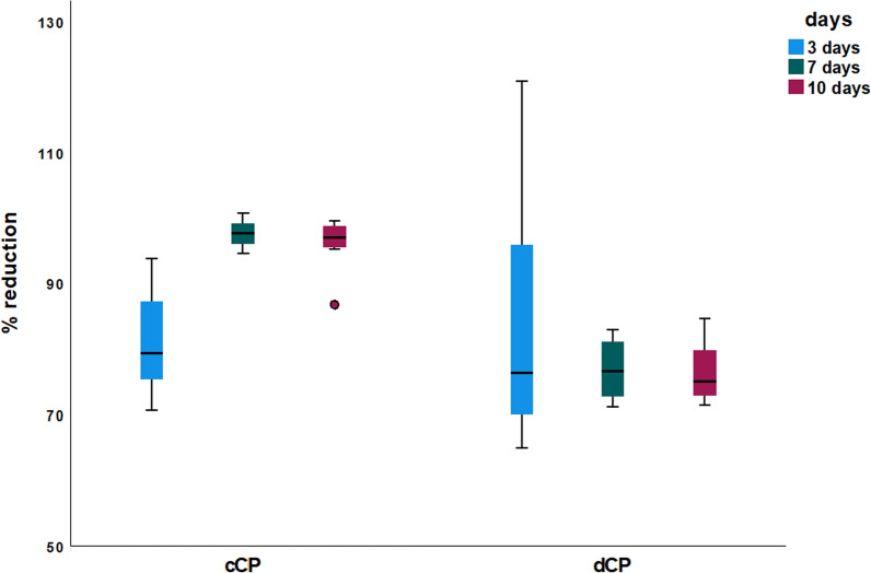
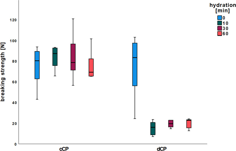
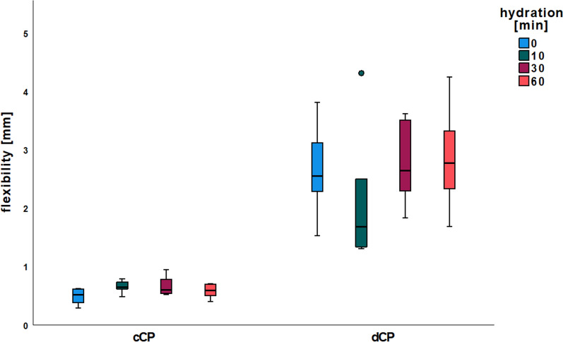
Methods: Breaking strength (BS; in N) and flexibility (F; in mm) of cCP and dCP were assessed and compared using four groups ((1) non-hydrated, (2) hydrated for 10, (3) 30, and (4) 60 min in saline), respectively. Cell viability of HOB was evaluated by resazurin reduction on non-hydrated cCP and dCP after 3, 7, and 10 days. Scanning electron microscopy (SEM) visualized CP breaking edges, internal structures, HOB cell morphology, and growth patterns.
Results: BS of hydrated dCP (d10: 15.45 ± 7.01 N, d30: 19.40 ± 3.78 N, d60: 20.31 ± 4.90 N) was significantly lower than that of non-hydrated dCP (d0: 74.70 ± 29.48 N) and native and hydrated cCP (c0: 75.00 ± 19.27 N, c10: 83.73 ± 10.92 N, c30: 83.80 ± 22.63 N, c60: 75.58 ± 14.25 N, p < 0.001 each). Next, dCP groups (d0: 2.64 ± 0.78 mm, d10: 2.14 ± 1.15 mm, d30: 2.76 ± 3.78 mm, d60: 2.86 ± 0.89 mm) exhibited significantly higher F than cCP groups (c0: 0.49 ± 0.14 mm, c10: 0.66 ± 0.10 mm, c30: 0.67 ± 0.16 mm, c60: 0.59 ± 0.12 mm, p < 0.05 each). No significant differences in F were observed among the different dCP groups. HOB cell viability was significantly increased on cCP compared to dCP after 7 (97.64 ± 2.11% vs. 76.88 ± 4.82%) and 10 days (96.14 ± 4.13% vs. 76.45 ± 4.64%; p < 0.001 each). SEM revealed well-defined breaking edges in cCP, whereas dCP displayed tear-off edges with shearing extensions. SEM showed disordered growth patterns and a physiological HOB cell morphology on dCP, contrasting with a parallel growth of fibroblast-like-looking HOB on cCP.
Conclusions: Compared to cCP, dCP showed increased flexibility but lower breaking strength and reduced HOB vitality. The increased flexibility and a decrease in breaking strength are likely due to differences in elasticity between dCP and cCP. The use of dCP may improve clinical handling efficiency.
目的:本研究旨在比较商业同种异体皮质骨板(cCP)和创新的不同脱矿CP (dCP)在生物力学和人成骨细胞(HOB)的体外和体外活力。方法:断裂强度(BS;N)和灵活性(F;采用四组((1)不补水,(2)生理盐水补水10分钟,(3)生理盐水补水30分钟,(4)生理盐水补水60分钟)对cCP和dCP进行评估和比较。3、7、10天后,用雷唑脲还原非水合cCP和dCP评价HOB细胞活力。扫描电子显微镜(SEM)显示了CP断裂边缘、内部结构、HOB细胞形态和生长模式。结果:水合dCP (d10: 15.45±7.01 N, d30: 19.40±3.78 N, d60: 20.31±4.90 N)的BS明显低于未水合dCP (d0: 74.70±29.48 N)和天然和水合cCP (c0: 75.00±19.27 N, c10: 83.73±10.92 N, c30: 83.80±22.63 N, c60: 75.58±14.25 N, p)。结论:与cCP相比,dCP的柔韧性增加,但断裂强度降低,HOB活力降低。韧性的增加和断裂强度的降低可能是由于dCP和cCP之间弹性的差异。使用dCP可提高临床处理效率。
期刊介绍:
The International Journal of Implant Dentistry is a peer-reviewed open access journal published under the SpringerOpen brand. The journal is dedicated to promoting the exchange and discussion of all research areas relevant to implant dentistry in the form of systematic literature or invited reviews, prospective and retrospective clinical studies, clinical case reports, basic laboratory and animal research, and articles on material research and engineering.
 求助内容:
求助内容: 应助结果提醒方式:


