Robert Abbott, Sarah Ward, Maya Jodidio, George Holan, Jeremy J Grachan
{"title":"Bilateral variations in the branching of the external carotid artery: a case report.","authors":"Robert Abbott, Sarah Ward, Maya Jodidio, George Holan, Jeremy J Grachan","doi":"10.1007/s00276-025-03653-5","DOIUrl":null,"url":null,"abstract":"<p><strong>Purpose: </strong>Various anatomical variations in the branching pattern of the external carotid arteries are known to occur with significant frequency and have been documented in published literature. The purpose of this case report is to document and discuss variations in the branching patterns of the external carotid artery as seen in an anatomical donor and determine the clinical relevance of these variations.</p><p><strong>Case presentation: </strong>A routine dissection of an 89-year-old female anatomical donor, whose cause of death was reported as acute myocardial infarction and atherosclerotic heart disease, revealed variations in the branching patterns of both external carotid arteries. Bilaterally, the common carotid arteries bifurcated at the C4 vertebral level. On the left side, an occipitoauricular trunk originated 0.5 mm superior to the common carotid artery's bifurcation, whereas, on the right side, an occipitoauriculopharyngeal trunk branching 0.8 mm superior to the bifurcation of the common carotid artery was observed before branching into an occipitoauricular trunk and ascending pharyngeal artery.</p><p><strong>Conclusion: </strong>This case report reinforces previous publications on arterial branching patterns and the importance of imaging prior to procedures. Clinically, these variations may impact surgical approaches, endovascular procedures within the neck, and vascular pathology management.</p>","PeriodicalId":49461,"journal":{"name":"Surgical and Radiologic Anatomy","volume":"47 1","pages":"141"},"PeriodicalIF":1.2000,"publicationDate":"2025-05-19","publicationTypes":"Journal Article","fieldsOfStudy":null,"isOpenAccess":false,"openAccessPdf":"https://www.ncbi.nlm.nih.gov/pmc/articles/PMC12089180/pdf/","citationCount":"0","resultStr":null,"platform":"Semanticscholar","paperid":null,"PeriodicalName":"Surgical and Radiologic Anatomy","FirstCategoryId":"3","ListUrlMain":"https://doi.org/10.1007/s00276-025-03653-5","RegionNum":4,"RegionCategory":"医学","ArticlePicture":[],"TitleCN":null,"AbstractTextCN":null,"PMCID":null,"EPubDate":"","PubModel":"","JCR":"Q2","JCRName":"Medicine","Score":null,"Total":0}
引用次数: 0
Abstract
Purpose: Various anatomical variations in the branching pattern of the external carotid arteries are known to occur with significant frequency and have been documented in published literature. The purpose of this case report is to document and discuss variations in the branching patterns of the external carotid artery as seen in an anatomical donor and determine the clinical relevance of these variations.
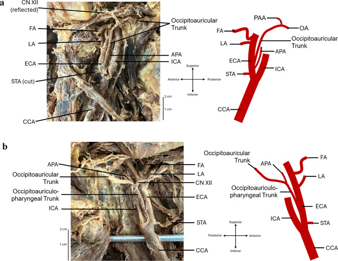
Case presentation: A routine dissection of an 89-year-old female anatomical donor, whose cause of death was reported as acute myocardial infarction and atherosclerotic heart disease, revealed variations in the branching patterns of both external carotid arteries. Bilaterally, the common carotid arteries bifurcated at the C4 vertebral level. On the left side, an occipitoauricular trunk originated 0.5 mm superior to the common carotid artery's bifurcation, whereas, on the right side, an occipitoauriculopharyngeal trunk branching 0.8 mm superior to the bifurcation of the common carotid artery was observed before branching into an occipitoauricular trunk and ascending pharyngeal artery.
Conclusion: This case report reinforces previous publications on arterial branching patterns and the importance of imaging prior to procedures. Clinically, these variations may impact surgical approaches, endovascular procedures within the neck, and vascular pathology management.
期刊介绍:
Anatomy is a morphological science which cannot fail to interest the clinician. The practical application of anatomical research to clinical problems necessitates special adaptation and selectivity in choosing from numerous international works. Although there is a tendency to believe that meaningful advances in anatomy are unlikely, constant revision is necessary. Surgical and Radiologic Anatomy, the first international journal of Clinical anatomy has been created in this spirit.
Its goal is to serve clinicians, regardless of speciality-physicians, surgeons, radiologists or other specialists-as an indispensable aid with which they can improve their knowledge of anatomy. Each issue includes: Original papers, review articles, articles on the anatomical bases of medical, surgical and radiological techniques, articles of normal radiologic anatomy, brief reviews of anatomical publications of clinical interest.
Particular attention is given to high quality illustrations, which are indispensable for a better understanding of anatomical problems.
Surgical and Radiologic Anatomy is a journal written by anatomists for clinicians with a special interest in anatomy.

 求助内容:
求助内容: 应助结果提醒方式:


