Eduardo Thadeu de Oliveira Correia, Jad Badreddine, Rasim Boyacioglu, Madison E Kretzler, Mark A Griswold, David Sheyn, Chris A Flask, Yong Chen, Adonis Hijaz, Leonardo Kayat Bittencourt
{"title":"Quantitative assessment of bladder tissue properties using magnetic resonance fingerprinting: a pilot feasibility study in healthy volunteers.","authors":"Eduardo Thadeu de Oliveira Correia, Jad Badreddine, Rasim Boyacioglu, Madison E Kretzler, Mark A Griswold, David Sheyn, Chris A Flask, Yong Chen, Adonis Hijaz, Leonardo Kayat Bittencourt","doi":"10.1590/0100-3984.2024.0104","DOIUrl":null,"url":null,"abstract":"<p><strong>Objective: </strong>To investigate the feasibility of performing magnetic resonance fingerprinting (MRF) of the bladder and quantify the T1 and T2 relaxation times of the bladder wall in healthy female volunteers, before and after voiding.</p><p><strong>Materials and methods: </strong>Volunteers without lower urinary tract symptoms underwent pelvic MRF. Five axial MRF slices of the bladder were obtained before and after voiding. Regions of interest were annotated on MRF T1 maps: one on the anterior bladder wall, and one on a lateral wall. Annotations made on T1 maps were subsequently copied to coregistered T2 maps. Student's t-tests for paired samples were employed to compare the T1 and T2 values obtained before voiding with those obtained after voiding.</p><p><strong>Results: </strong>Eight volunteers were included. The mean preand post-void T1 relaxation times were 1,575 ± 93 ms and 1,476 ± 138 ms, respectively. The mean preand post-void T2 relaxation times were 55 ± 21 ms and 53 ± 8 ms, respectively. The mean T1 relaxation times were 6% lower after voiding than before (<i>p</i> = 0.035).</p><p><strong>Conclusion: </strong>The use of MRF to quantify T1 and T2 relaxation times in the bladder appears to be feasible. Our results can serve as a reference for studies investigating T1 and T2 relaxation times in patients with malignant or nonmalignant bladder disorders.</p>","PeriodicalId":20842,"journal":{"name":"Radiologia Brasileira","volume":"58 ","pages":"e20240104"},"PeriodicalIF":0.0000,"publicationDate":"2025-05-01","publicationTypes":"Journal Article","fieldsOfStudy":null,"isOpenAccess":false,"openAccessPdf":"https://www.ncbi.nlm.nih.gov/pmc/articles/PMC12087349/pdf/","citationCount":"0","resultStr":null,"platform":"Semanticscholar","paperid":null,"PeriodicalName":"Radiologia Brasileira","FirstCategoryId":"1085","ListUrlMain":"https://doi.org/10.1590/0100-3984.2024.0104","RegionNum":0,"RegionCategory":null,"ArticlePicture":[],"TitleCN":null,"AbstractTextCN":null,"PMCID":null,"EPubDate":"2025/1/1 0:00:00","PubModel":"eCollection","JCR":"Q3","JCRName":"Medicine","Score":null,"Total":0}
引用次数: 0
Abstract
Objective: To investigate the feasibility of performing magnetic resonance fingerprinting (MRF) of the bladder and quantify the T1 and T2 relaxation times of the bladder wall in healthy female volunteers, before and after voiding.
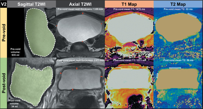
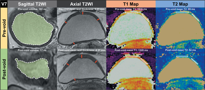
Materials and methods: Volunteers without lower urinary tract symptoms underwent pelvic MRF. Five axial MRF slices of the bladder were obtained before and after voiding. Regions of interest were annotated on MRF T1 maps: one on the anterior bladder wall, and one on a lateral wall. Annotations made on T1 maps were subsequently copied to coregistered T2 maps. Student's t-tests for paired samples were employed to compare the T1 and T2 values obtained before voiding with those obtained after voiding.
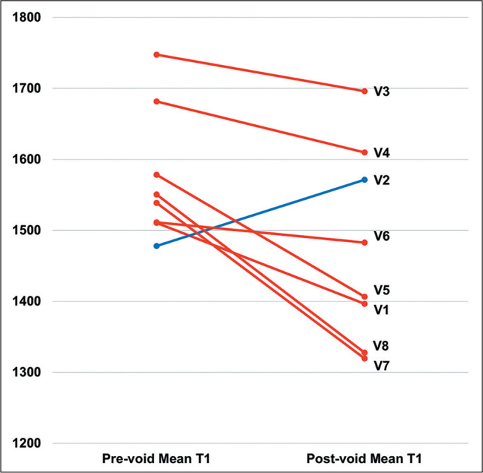
Results: Eight volunteers were included. The mean preand post-void T1 relaxation times were 1,575 ± 93 ms and 1,476 ± 138 ms, respectively. The mean preand post-void T2 relaxation times were 55 ± 21 ms and 53 ± 8 ms, respectively. The mean T1 relaxation times were 6% lower after voiding than before (p = 0.035).
Conclusion: The use of MRF to quantify T1 and T2 relaxation times in the bladder appears to be feasible. Our results can serve as a reference for studies investigating T1 and T2 relaxation times in patients with malignant or nonmalignant bladder disorders.

 求助内容:
求助内容: 应助结果提醒方式:


