{"title":"[Role of radiology for primary staging of colorectal cancer].","authors":"Sophia Wirth, Nino Bogveradze, Katharina Lampichler","doi":"10.1007/s00117-025-01458-6","DOIUrl":null,"url":null,"abstract":"<p><strong>Background: </strong>Imaging, specifically magnetic resonance imaging (MRI), has long been the cornerstone for staging rectal cancer. In contrast, computed tomography (CT) is the standard for colon cancer staging. Primary staging is essential for individualized risk stratification and therapy planning.</p><p><strong>Objectives: </strong>To summarize current international guidelines on primary colorectal cancer staging, taking into account the advantages and disadvantages of different imaging methods.</p><p><strong>Materials and methods: </strong>Based on extensive literature research, the current state of knowledge in the staging of colorectal cancer is summarized.</p><p><strong>Results: </strong>TNM staging includes local tumor extension as well as the presence of lymph node and distant metastases. For local tumor extension, MRI is very well suited for rectal cancer. In colon cancer, CT shows significant limitations regarding the sensitivity of differentiating individual stages. Lymph node metastases can pose a major challenge for both MRI and CT, which has led to the definition of several malignancy criteria. The detection of distant metastases remains primarily within the domain of CT, except for liver metastases.</p><p><strong>Conclusions: </strong>Depending on tumor location, either MRI or CT is recommended for primary staging of colorectal cancer, and a combination of both modalities may also be appropriate. Accurate assessment of the primary tumor, lymph nodes, and organs using imaging is an essential component of the therapeutic approach to colorectal cancer.</p>","PeriodicalId":74635,"journal":{"name":"Radiologie (Heidelberg, Germany)","volume":" ","pages":"426-435"},"PeriodicalIF":0.6000,"publicationDate":"2025-06-01","publicationTypes":"Journal Article","fieldsOfStudy":null,"isOpenAccess":false,"openAccessPdf":"https://www.ncbi.nlm.nih.gov/pmc/articles/PMC12106153/pdf/","citationCount":"0","resultStr":null,"platform":"Semanticscholar","paperid":null,"PeriodicalName":"Radiologie (Heidelberg, Germany)","FirstCategoryId":"1085","ListUrlMain":"https://doi.org/10.1007/s00117-025-01458-6","RegionNum":0,"RegionCategory":null,"ArticlePicture":[],"TitleCN":null,"AbstractTextCN":null,"PMCID":null,"EPubDate":"2025/5/19 0:00:00","PubModel":"Epub","JCR":"","JCRName":"","Score":null,"Total":0}
引用次数: 0
Abstract
Background: Imaging, specifically magnetic resonance imaging (MRI), has long been the cornerstone for staging rectal cancer. In contrast, computed tomography (CT) is the standard for colon cancer staging. Primary staging is essential for individualized risk stratification and therapy planning.
Objectives: To summarize current international guidelines on primary colorectal cancer staging, taking into account the advantages and disadvantages of different imaging methods.
Materials and methods: Based on extensive literature research, the current state of knowledge in the staging of colorectal cancer is summarized.
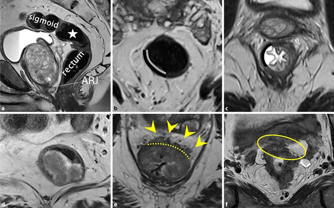
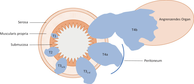
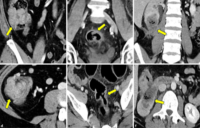
Results: TNM staging includes local tumor extension as well as the presence of lymph node and distant metastases. For local tumor extension, MRI is very well suited for rectal cancer. In colon cancer, CT shows significant limitations regarding the sensitivity of differentiating individual stages. Lymph node metastases can pose a major challenge for both MRI and CT, which has led to the definition of several malignancy criteria. The detection of distant metastases remains primarily within the domain of CT, except for liver metastases.
Conclusions: Depending on tumor location, either MRI or CT is recommended for primary staging of colorectal cancer, and a combination of both modalities may also be appropriate. Accurate assessment of the primary tumor, lymph nodes, and organs using imaging is an essential component of the therapeutic approach to colorectal cancer.

 求助内容:
求助内容: 应助结果提醒方式:


