{"title":"Morphometric and radiomics analysis toward the prediction of epilepsy associated with supratentorial low-grade glioma in children.","authors":"Min-Lan Tsai, Kevin Li-Chun Hsieh, Yen-Lin Liu, Yi-Shan Yang, Hsi Chang, Tai-Tong Wong, Syu-Jyun Peng","doi":"10.1186/s40644-025-00881-1","DOIUrl":null,"url":null,"abstract":"<p><strong>Objectives: </strong>Understanding the impact of epilepsy on pediatric brain tumors is crucial to diagnostic precision and optimal treatment selection. This study investigated MRI radiomics features, tumor location, voxel-based morphometry (VBM) for gray matter density, and tumor volumetry to differentiate between children with low grade glioma (LGG)-associated epilepsies and those without, and further identified key radiomics features for predicting of epilepsy risk in children with supratentorial LGG to construct an epilepsy prediction model.</p><p><strong>Methods: </strong>A total of 206 radiomics features of tumors and voxel-based morphometric analysis of tumor location features were extracted from T2-FLAIR images in a primary cohort of 48 children with LGG with epilepsy (N = 23) or without epilepsy (N = 25), prior to surgery. Feature selection was performed using the minimum redundancy maximum relevance algorithm, and leave-one-out cross-validation was applied to assess the predictive performance of radiomics and tumor location signatures in differentiating epilepsy-associated LGG from non-epilepsy cases.</p><p><strong>Results: </strong>Voxel-based morphometric analysis showed significant positive t-scores within bilateral temporal cortex and negative t-scores in basal ganglia between epilepsy and non-epilepsy groups. Eight radiomics features were identified as significant predictors of epilepsy in LGG, encompassing characteristics of 2 locations, 2 shapes, 1 image gray scale intensity, and 3 textures. The most important predictor was temporal lobe involvement, followed by high dependence high grey level emphasis, elongation, area density, information correlation 1, midbrain and intensity range. The Linear Support Vector Machine (SVM) model yielded the best prediction performance, when implemented with a combination of radiomics features and tumor location features, as evidenced by the following metrics: precision (0.955), recall (0.913), specificity (0.960), accuracy (0.938), F-1 score (0.933), and area under curve (AUC) (0.950).</p><p><strong>Conclusion: </strong>Our findings demonstrated the efficacy of machine learning models based on radiomics features and voxel-based anatomical locations in predicting the risk of epilepsy in supratentorial LGG. This model provides a highly accurate tool for distinguishing epilepsy-associated LGG in children, supporting precise treatment planning.</p><p><strong>Trial registration: </strong>Not applicable.</p>","PeriodicalId":9548,"journal":{"name":"Cancer Imaging","volume":"25 1","pages":"63"},"PeriodicalIF":3.5000,"publicationDate":"2025-05-19","publicationTypes":"Journal Article","fieldsOfStudy":null,"isOpenAccess":false,"openAccessPdf":"https://www.ncbi.nlm.nih.gov/pmc/articles/PMC12090388/pdf/","citationCount":"0","resultStr":null,"platform":"Semanticscholar","paperid":null,"PeriodicalName":"Cancer Imaging","FirstCategoryId":"3","ListUrlMain":"https://doi.org/10.1186/s40644-025-00881-1","RegionNum":2,"RegionCategory":"医学","ArticlePicture":[],"TitleCN":null,"AbstractTextCN":null,"PMCID":null,"EPubDate":"","PubModel":"","JCR":"Q2","JCRName":"ONCOLOGY","Score":null,"Total":0}
引用次数: 0
Abstract
Objectives: Understanding the impact of epilepsy on pediatric brain tumors is crucial to diagnostic precision and optimal treatment selection. This study investigated MRI radiomics features, tumor location, voxel-based morphometry (VBM) for gray matter density, and tumor volumetry to differentiate between children with low grade glioma (LGG)-associated epilepsies and those without, and further identified key radiomics features for predicting of epilepsy risk in children with supratentorial LGG to construct an epilepsy prediction model.
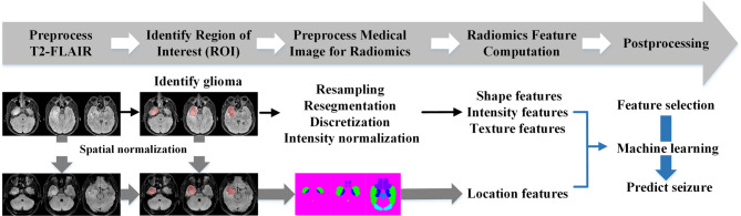
Methods: A total of 206 radiomics features of tumors and voxel-based morphometric analysis of tumor location features were extracted from T2-FLAIR images in a primary cohort of 48 children with LGG with epilepsy (N = 23) or without epilepsy (N = 25), prior to surgery. Feature selection was performed using the minimum redundancy maximum relevance algorithm, and leave-one-out cross-validation was applied to assess the predictive performance of radiomics and tumor location signatures in differentiating epilepsy-associated LGG from non-epilepsy cases.
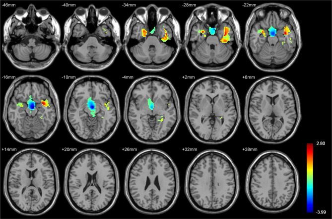
Results: Voxel-based morphometric analysis showed significant positive t-scores within bilateral temporal cortex and negative t-scores in basal ganglia between epilepsy and non-epilepsy groups. Eight radiomics features were identified as significant predictors of epilepsy in LGG, encompassing characteristics of 2 locations, 2 shapes, 1 image gray scale intensity, and 3 textures. The most important predictor was temporal lobe involvement, followed by high dependence high grey level emphasis, elongation, area density, information correlation 1, midbrain and intensity range. The Linear Support Vector Machine (SVM) model yielded the best prediction performance, when implemented with a combination of radiomics features and tumor location features, as evidenced by the following metrics: precision (0.955), recall (0.913), specificity (0.960), accuracy (0.938), F-1 score (0.933), and area under curve (AUC) (0.950).
Conclusion: Our findings demonstrated the efficacy of machine learning models based on radiomics features and voxel-based anatomical locations in predicting the risk of epilepsy in supratentorial LGG. This model provides a highly accurate tool for distinguishing epilepsy-associated LGG in children, supporting precise treatment planning.
Cancer ImagingONCOLOGY-RADIOLOGY, NUCLEAR MEDICINE & MEDICAL IMAGING
CiteScore
7.00
自引率
0.00%
发文量
66
审稿时长
>12 weeks
期刊介绍:
Cancer Imaging is an open access, peer-reviewed journal publishing original articles, reviews and editorials written by expert international radiologists working in oncology.
The journal encompasses CT, MR, PET, ultrasound, radionuclide and multimodal imaging in all kinds of malignant tumours, plus new developments, techniques and innovations. Topics of interest include:
Breast Imaging
Chest
Complications of treatment
Ear, Nose & Throat
Gastrointestinal
Hepatobiliary & Pancreatic
Imaging biomarkers
Interventional
Lymphoma
Measurement of tumour response
Molecular functional imaging
Musculoskeletal
Neuro oncology
Nuclear Medicine
Paediatric.

 求助内容:
求助内容: 应助结果提醒方式:


