I F Gosselink, P Leonhardt, E M Höppener, R Smelt, M J Drittij, M Davigo, G G H van den Akker, I M Kooter, T J M Welting, F J van Schooten, A H V Remels
{"title":"Size- and polymer-dependent toxicity of amorphous environmentally relevant micro- and nanoplastics in human bronchial epithelial cells.","authors":"I F Gosselink, P Leonhardt, E M Höppener, R Smelt, M J Drittij, M Davigo, G G H van den Akker, I M Kooter, T J M Welting, F J van Schooten, A H V Remels","doi":"10.1186/s43591-025-00126-9","DOIUrl":null,"url":null,"abstract":"<p><strong>Background: </strong>Knowledge of the toxicological impact of micro- and nanoplastics (MNPs) on the human airway epithelium is limited and almost exclusively based on experiments applying high doses of spherical polystyrene (PS) particles. In this study, we investigated the toxicity of a broad size range of amorphous MNPs generated from different environmentally-relevant polymers.</p><p><strong>Methods: </strong>Bronchial epithelial cells (BEAS-2B) were exposed to three different doses of polyvinylchloride (PVC), polypropylene (PP), or polyamide (PA) particles (< 1 μm-10 μm), as well as leachates from these polymers. Toxicity was evaluated by assessment of cytotoxicity, inflammation (IL-8 release and inflammatory gene expression) and oxidative stress (DCFH-DA assay and antioxidant gene expression). Furthermore, the molecular mechanism behind MNP-induced inflammation was investigated by studying activation of two well-known inflammation related transcriptional factors (NF-κB and AP-1).</p><p><strong>Results: </strong>Only PA nanoplastics induced significant cell death, IL-8 secretion and inflammatory gene expression compared to vehicle control. PA-induced inflammation was accompanied by NF-κB, but not AP-1, transcriptional activity. PA did not increase cellular ROS levels; however, it did lead to increased expression of the antioxidant gene superoxide dismutase 2. In addition to PA, exposure to < 1 µm and 1-5 µm PP particles resulted in elevated IL-8 secretion, likely due to the presence of talc added as filler. None of the leachates affected cytotoxicity or inflammation.</p><p><strong>Conclusion: </strong>Toxicity of MNPs to human bronchial epithelial cells was dependent on polymer type, size and dose. Nanoplastics, especially PA, were more toxic to bronchial epithelial cells than microplastics and induced cytotoxicity and an inflammatory response.</p><p><strong>Graphical abstract: </strong></p><p><strong>Supplementary information: </strong>The online version contains supplementary material available at 10.1186/s43591-025-00126-9.</p>","PeriodicalId":74190,"journal":{"name":"Microplastics and nanoplastics","volume":"5 1","pages":"19"},"PeriodicalIF":0.0000,"publicationDate":"2025-01-01","publicationTypes":"Journal Article","fieldsOfStudy":null,"isOpenAccess":false,"openAccessPdf":"https://www.ncbi.nlm.nih.gov/pmc/articles/PMC12081513/pdf/","citationCount":"0","resultStr":null,"platform":"Semanticscholar","paperid":null,"PeriodicalName":"Microplastics and nanoplastics","FirstCategoryId":"1085","ListUrlMain":"https://doi.org/10.1186/s43591-025-00126-9","RegionNum":0,"RegionCategory":null,"ArticlePicture":[],"TitleCN":null,"AbstractTextCN":null,"PMCID":null,"EPubDate":"2025/5/16 0:00:00","PubModel":"Epub","JCR":"","JCRName":"","Score":null,"Total":0}
引用次数: 0
Abstract
Background: Knowledge of the toxicological impact of micro- and nanoplastics (MNPs) on the human airway epithelium is limited and almost exclusively based on experiments applying high doses of spherical polystyrene (PS) particles. In this study, we investigated the toxicity of a broad size range of amorphous MNPs generated from different environmentally-relevant polymers.
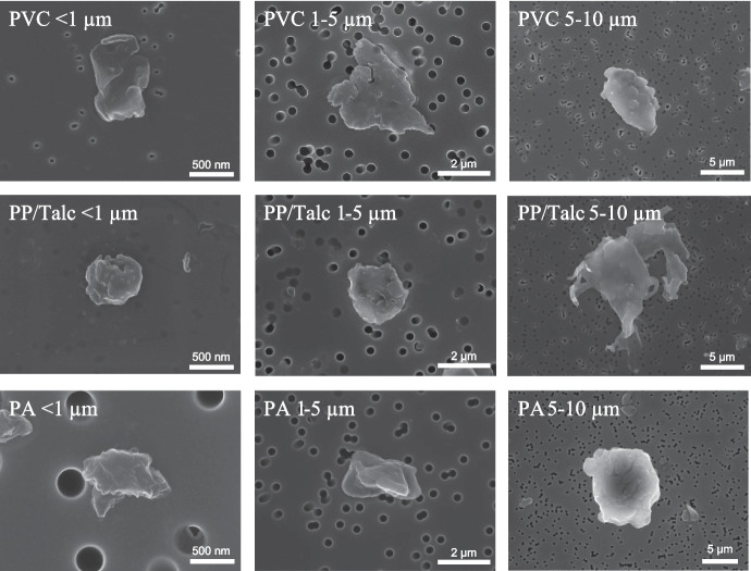
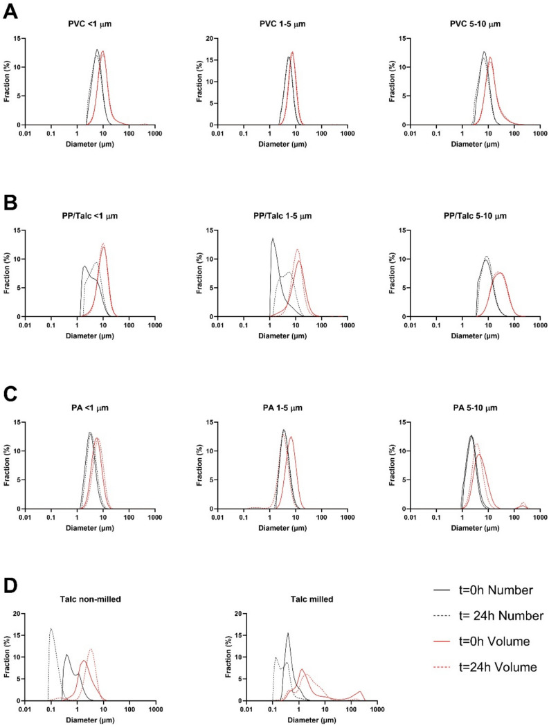
Methods: Bronchial epithelial cells (BEAS-2B) were exposed to three different doses of polyvinylchloride (PVC), polypropylene (PP), or polyamide (PA) particles (< 1 μm-10 μm), as well as leachates from these polymers. Toxicity was evaluated by assessment of cytotoxicity, inflammation (IL-8 release and inflammatory gene expression) and oxidative stress (DCFH-DA assay and antioxidant gene expression). Furthermore, the molecular mechanism behind MNP-induced inflammation was investigated by studying activation of two well-known inflammation related transcriptional factors (NF-κB and AP-1).
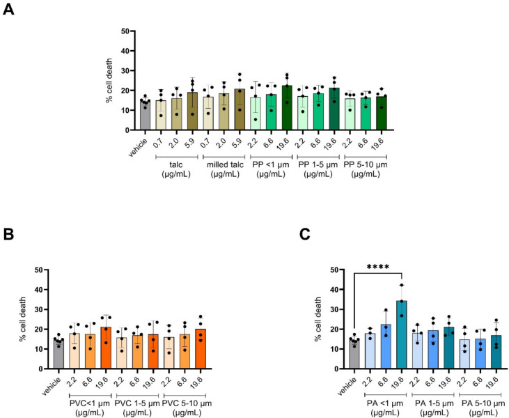
Results: Only PA nanoplastics induced significant cell death, IL-8 secretion and inflammatory gene expression compared to vehicle control. PA-induced inflammation was accompanied by NF-κB, but not AP-1, transcriptional activity. PA did not increase cellular ROS levels; however, it did lead to increased expression of the antioxidant gene superoxide dismutase 2. In addition to PA, exposure to < 1 µm and 1-5 µm PP particles resulted in elevated IL-8 secretion, likely due to the presence of talc added as filler. None of the leachates affected cytotoxicity or inflammation.
Conclusion: Toxicity of MNPs to human bronchial epithelial cells was dependent on polymer type, size and dose. Nanoplastics, especially PA, were more toxic to bronchial epithelial cells than microplastics and induced cytotoxicity and an inflammatory response.
Graphical abstract:
Supplementary information: The online version contains supplementary material available at 10.1186/s43591-025-00126-9.

 求助内容:
求助内容: 应助结果提醒方式:


