{"title":"PET/CT Radiomics Integrated with Clinical Indexes as a Tool to Predict Ki67 in Breast Cancer: a Pilot Study.","authors":"Dawei Li, Hui Ding, Yuting Liao, Xiao Yu, Youmin Guo, Cong Shen","doi":"10.1007/s13139-024-00896-9","DOIUrl":null,"url":null,"abstract":"<p><strong>Objective: </strong>This study aims to assess the value of radiomics features integrated with clinical characteristics for estimating Ki67 expression in patients with breast cancer (BC).</p><p><strong>Methods: </strong>In total, 114 patients with BC performed <sup>18</sup>F-FDG PET/CT scans. Patients were randomly assigned to a training set (<i>n</i> = 79, 55 cases of Ki67 + and 24 cases of Ki67-) and a validation set (<i>n</i> = 35, 24 cases of Ki67 + and 11 cases of Ki67-). Thirteen clinical characteristics and 704 radiomics features were extracted, and 4 clinical and 8 radiomics features were selected. Three models were developed, including the clinical model, the radiomics model, and the combined model. Model performance was evaluated using the ROC curve, and clinical utility was assessed through decision curve analysis (DCA).</p><p><strong>Results: </strong>The N stage, tumor morphology, SUVmax, and the longest diameter significantly differed between Ki67 + and Ki67- groups (all <i>P</i> < 0.05). Eight radiomics features were selected for the radiomics model. The area under the curve of the combined model in the training and test group was 0.90 (95% CI: 0.82∼0.97) and 0.81 (95% CI: 0.64∼0.99), respectively. The combined model significantly outperformed both the radiomics model and the clinical model alone (<i>P</i> < 0.05). The DCA curve demonstrated the superior clinical utility of the combined model compared to the clinical model and radiomics model.</p><p><strong>Conclusions: </strong>PET/CT image-based radiomics features combined with clinical features have the potential to predict Ki67 expression in BC.</p><p><strong>Supplementary information: </strong>The online version contains supplementary material available at 10.1007/s13139-024-00896-9.</p>","PeriodicalId":19384,"journal":{"name":"Nuclear Medicine and Molecular Imaging","volume":"59 3","pages":"164-173"},"PeriodicalIF":2.7000,"publicationDate":"2025-06-01","publicationTypes":"Journal Article","fieldsOfStudy":null,"isOpenAccess":false,"openAccessPdf":"https://www.ncbi.nlm.nih.gov/pmc/articles/PMC12084439/pdf/","citationCount":"0","resultStr":null,"platform":"Semanticscholar","paperid":null,"PeriodicalName":"Nuclear Medicine and Molecular Imaging","FirstCategoryId":"1085","ListUrlMain":"https://doi.org/10.1007/s13139-024-00896-9","RegionNum":0,"RegionCategory":null,"ArticlePicture":[],"TitleCN":null,"AbstractTextCN":null,"PMCID":null,"EPubDate":"2024/11/29 0:00:00","PubModel":"Epub","JCR":"Q3","JCRName":"RADIOLOGY, NUCLEAR MEDICINE & MEDICAL IMAGING","Score":null,"Total":0}
引用次数: 0
Abstract
Objective: This study aims to assess the value of radiomics features integrated with clinical characteristics for estimating Ki67 expression in patients with breast cancer (BC).
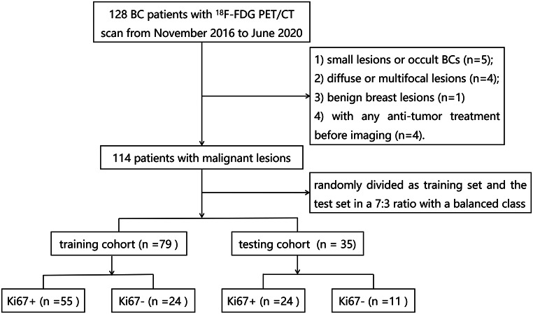
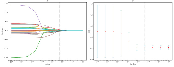
Methods: In total, 114 patients with BC performed 18F-FDG PET/CT scans. Patients were randomly assigned to a training set (n = 79, 55 cases of Ki67 + and 24 cases of Ki67-) and a validation set (n = 35, 24 cases of Ki67 + and 11 cases of Ki67-). Thirteen clinical characteristics and 704 radiomics features were extracted, and 4 clinical and 8 radiomics features were selected. Three models were developed, including the clinical model, the radiomics model, and the combined model. Model performance was evaluated using the ROC curve, and clinical utility was assessed through decision curve analysis (DCA).
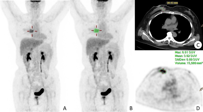
Results: The N stage, tumor morphology, SUVmax, and the longest diameter significantly differed between Ki67 + and Ki67- groups (all P < 0.05). Eight radiomics features were selected for the radiomics model. The area under the curve of the combined model in the training and test group was 0.90 (95% CI: 0.82∼0.97) and 0.81 (95% CI: 0.64∼0.99), respectively. The combined model significantly outperformed both the radiomics model and the clinical model alone (P < 0.05). The DCA curve demonstrated the superior clinical utility of the combined model compared to the clinical model and radiomics model.
Conclusions: PET/CT image-based radiomics features combined with clinical features have the potential to predict Ki67 expression in BC.
Supplementary information: The online version contains supplementary material available at 10.1007/s13139-024-00896-9.
期刊介绍:
Nuclear Medicine and Molecular Imaging (Nucl Med Mol Imaging) is an official journal of the Korean Society of Nuclear Medicine, which bimonthly publishes papers on February, April, June, August, October, and December about nuclear medicine and related sciences such as radiochemistry, radiopharmacy, dosimetry and pharmacokinetics / pharmacodynamics of radiopharmaceuticals, nuclear and molecular imaging analysis, nuclear and molecular imaging instrumentation, radiation biology and radionuclide therapy. The journal specially welcomes works of artificial intelligence applied to nuclear medicine. The journal will also welcome original works relating to molecular imaging research such as the development of molecular imaging probes, reporter imaging assays, imaging cell trafficking, imaging endo(exo)genous gene expression, and imaging signal transduction. Nucl Med Mol Imaging publishes the following types of papers: original articles, reviews, case reports, editorials, interesting images, and letters to the editor.
The Korean Society of Nuclear Medicine (KSNM)
KSNM is a scientific and professional organization founded in 1961 and a member of the Korean Academy of Medical Sciences of the Korean Medical Association which was established by The Medical Services Law. The aims of KSNM are the promotion of nuclear medicine and cooperation of each member. The business of KSNM includes holding academic meetings and symposia, the publication of journals and books, planning and research of promoting science and health, and training and qualification of nuclear medicine specialists.
 求助内容:
求助内容: 应助结果提醒方式:


