Jonas Biehler, Marie Brei, Nina Pischke, Sebastian Rasch, Miriam Dibos, Johanna Erber, Roland M Schmid, Rickmer F Braren, Markus R Makowski, Karl-Robert Wichmann, Kei Wieland Mueller, Wolfgang A Wall, Tobias Lahmer
{"title":"Approximation of EVLWI in severe COVID-19 pneumonia using quantitative imaging techniques: an observational study.","authors":"Jonas Biehler, Marie Brei, Nina Pischke, Sebastian Rasch, Miriam Dibos, Johanna Erber, Roland M Schmid, Rickmer F Braren, Markus R Makowski, Karl-Robert Wichmann, Kei Wieland Mueller, Wolfgang A Wall, Tobias Lahmer","doi":"10.1186/s40635-025-00752-w","DOIUrl":null,"url":null,"abstract":"<p><strong>Background: </strong>This study aimed to approximate the level of extravascular lung water (EVLW) in patients with severe COVID-19 pneumonia using quantitative imaging techniques. The elevation of EVLW is known to correlate with the degree of diffuse alveolar damage and linked with the mortality of critically ill patients. Transpulmonary thermodilution (TPTD) is the gold standard technique to estimate the total amount of EVLW, but it is invasive and requires specialized equipment and trained personnel.</p><p><strong>Methods: </strong>The study included patients with severe COVID-19 who required chest CT scanning within the first 48 h of Intensive Care Unit (ICU) admission and had TPTD monitoring. Using in-house software tools for automatic semantic segmentation, lung masks were obtained for estimating the EVLW content. The results were compared with the TPTD measurements.</p><p><strong>Results: </strong>The results demonstrate a significant correlation between EVLW-TPTP measured by thermodilution and EVLW-CT estimated from the patient's CT-image (r = 0.629, p = 0.0014).</p><p><strong>Conclusion: </strong>The study showed that quantitative imaging techniques using chest CT-scans could be used as a convenient and low-cost option for ICUs without TPTD equipment for the assessment of EVLW in severe COVID-19 pneumonia.</p>","PeriodicalId":13750,"journal":{"name":"Intensive Care Medicine Experimental","volume":"13 1","pages":"52"},"PeriodicalIF":2.8000,"publicationDate":"2025-05-19","publicationTypes":"Journal Article","fieldsOfStudy":null,"isOpenAccess":false,"openAccessPdf":"https://www.ncbi.nlm.nih.gov/pmc/articles/PMC12089548/pdf/","citationCount":"0","resultStr":null,"platform":"Semanticscholar","paperid":null,"PeriodicalName":"Intensive Care Medicine Experimental","FirstCategoryId":"1085","ListUrlMain":"https://doi.org/10.1186/s40635-025-00752-w","RegionNum":0,"RegionCategory":null,"ArticlePicture":[],"TitleCN":null,"AbstractTextCN":null,"PMCID":null,"EPubDate":"","PubModel":"","JCR":"Q2","JCRName":"CRITICAL CARE MEDICINE","Score":null,"Total":0}
引用次数: 0
Abstract
Background: This study aimed to approximate the level of extravascular lung water (EVLW) in patients with severe COVID-19 pneumonia using quantitative imaging techniques. The elevation of EVLW is known to correlate with the degree of diffuse alveolar damage and linked with the mortality of critically ill patients. Transpulmonary thermodilution (TPTD) is the gold standard technique to estimate the total amount of EVLW, but it is invasive and requires specialized equipment and trained personnel.
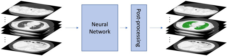
Methods: The study included patients with severe COVID-19 who required chest CT scanning within the first 48 h of Intensive Care Unit (ICU) admission and had TPTD monitoring. Using in-house software tools for automatic semantic segmentation, lung masks were obtained for estimating the EVLW content. The results were compared with the TPTD measurements.
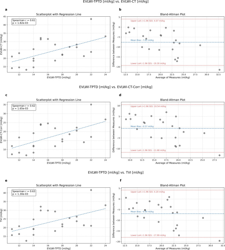
Results: The results demonstrate a significant correlation between EVLW-TPTP measured by thermodilution and EVLW-CT estimated from the patient's CT-image (r = 0.629, p = 0.0014).
Conclusion: The study showed that quantitative imaging techniques using chest CT-scans could be used as a convenient and low-cost option for ICUs without TPTD equipment for the assessment of EVLW in severe COVID-19 pneumonia.

 求助内容:
求助内容: 应助结果提醒方式:


