The role of artificial intelligence and deep learning in determining the histopathological grade of pancreatic neuroendocrine tumors by using EUS images.
{"title":"The role of artificial intelligence and deep learning in determining the histopathological grade of pancreatic neuroendocrine tumors by using EUS images.","authors":"Sercan Kiremitci, Gulseren Seven, Gokhan Silahtaroglu, Koray Kochan, Serife Degirmencioglu Tosun, Hakan Senturk","doi":"10.1097/eus.0000000000000113","DOIUrl":null,"url":null,"abstract":"<p><strong>Background and objectives: </strong>Pancreatic neuroendocrine tumors (pNETs) are relatively rare and consist of 2% of the all pancreatic tumors. Although some of pNETs have a benign, nonprogressive course, some may be progressive and result with metastasis. We aimed to estimate the grade of pNETs by using artificial intelligence (AI) via deep learning (DL) algorithms as indexing to cyto/histopathological classification according to the World Health Organization 2017.</p><p><strong>Methods: </strong>A total of 803 EUS images were collected from 44 patients who had a cyto/histo-pathologically confirmed diagnosis with EUS fine-needle aspiration or biopsy (FNA/B). First, raw EUS images were prepared for processing by AI via DL algorithms, and convolutional neural networks were utilized to train the machine to predict the grades from EUS images. IBM SPSS 25.0 program was used for statistical analyses.</p><p><strong>Results: </strong>Thirty of the 44 patients (68%) were female, with a median age of 61 (range, 16-80) years. pNETs were mostly located in the pancreatic head: 24 cases (55%). Location was the neck in 3 (7%), body in 10 (22%), and tail in 7 (16%) patients. According to EUS-FNA/B results, 27 patients were grade 1 (G1) (61%); 12, grade 2 (G2) (27%); and 5, grade 3 (G3) (12%). In reference to the performance of AI for predicting the pathological grade, sensitivity was 94.29%; specificity, 97.14%; and accuracy, 96.19%. When the patient groups were subanalyzed as G1, G2, and G3 by the AI model to predict the pathological grade, the accuracy was as follows: for G1, 93.15%; for G2, 91.61%; and for G3, 98.05%.</p><p><strong>Conclusions: </strong>This pilot study suggests that pNET grade prediction can be reliably done on EUS images using AI-based technology.</p>","PeriodicalId":11577,"journal":{"name":"Endoscopic Ultrasound","volume":"14 2","pages":"48-56"},"PeriodicalIF":5.4000,"publicationDate":"2025-03-01","publicationTypes":"Journal Article","fieldsOfStudy":null,"isOpenAccess":false,"openAccessPdf":"https://www.ncbi.nlm.nih.gov/pmc/articles/PMC12080687/pdf/","citationCount":"0","resultStr":null,"platform":"Semanticscholar","paperid":null,"PeriodicalName":"Endoscopic Ultrasound","FirstCategoryId":"3","ListUrlMain":"https://doi.org/10.1097/eus.0000000000000113","RegionNum":1,"RegionCategory":"医学","ArticlePicture":[],"TitleCN":null,"AbstractTextCN":null,"PMCID":null,"EPubDate":"2025/5/2 0:00:00","PubModel":"Epub","JCR":"Q1","JCRName":"GASTROENTEROLOGY & HEPATOLOGY","Score":null,"Total":0}
引用次数: 0
Abstract
Background and objectives: Pancreatic neuroendocrine tumors (pNETs) are relatively rare and consist of 2% of the all pancreatic tumors. Although some of pNETs have a benign, nonprogressive course, some may be progressive and result with metastasis. We aimed to estimate the grade of pNETs by using artificial intelligence (AI) via deep learning (DL) algorithms as indexing to cyto/histopathological classification according to the World Health Organization 2017.
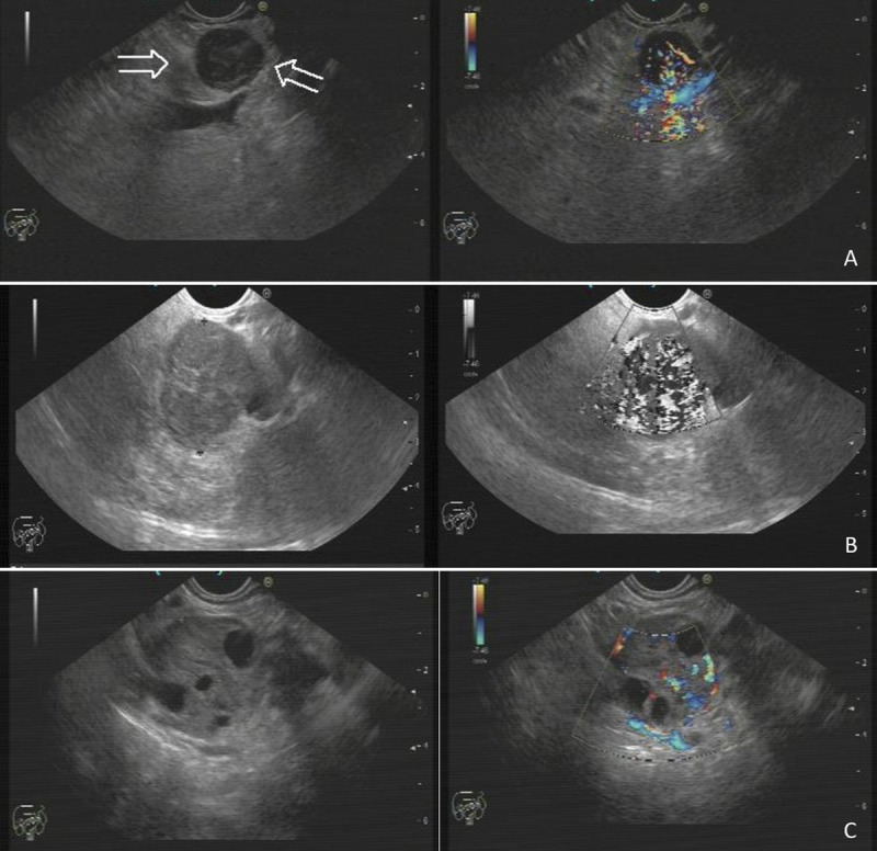
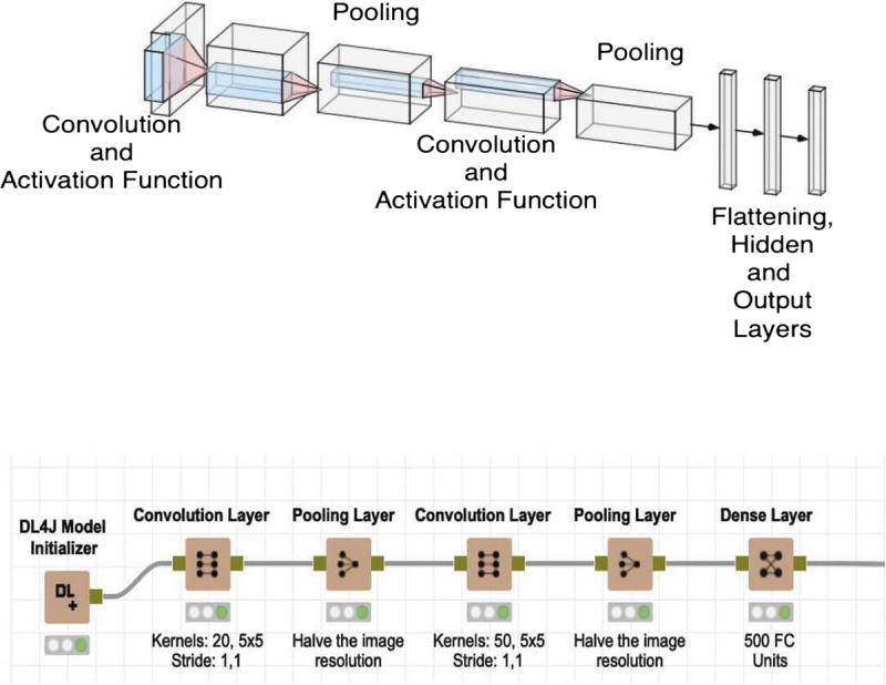
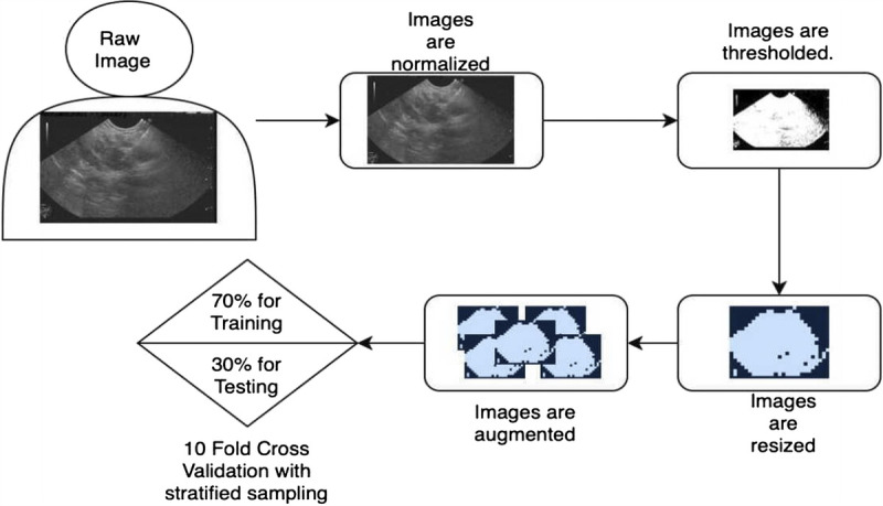
Methods: A total of 803 EUS images were collected from 44 patients who had a cyto/histo-pathologically confirmed diagnosis with EUS fine-needle aspiration or biopsy (FNA/B). First, raw EUS images were prepared for processing by AI via DL algorithms, and convolutional neural networks were utilized to train the machine to predict the grades from EUS images. IBM SPSS 25.0 program was used for statistical analyses.
Results: Thirty of the 44 patients (68%) were female, with a median age of 61 (range, 16-80) years. pNETs were mostly located in the pancreatic head: 24 cases (55%). Location was the neck in 3 (7%), body in 10 (22%), and tail in 7 (16%) patients. According to EUS-FNA/B results, 27 patients were grade 1 (G1) (61%); 12, grade 2 (G2) (27%); and 5, grade 3 (G3) (12%). In reference to the performance of AI for predicting the pathological grade, sensitivity was 94.29%; specificity, 97.14%; and accuracy, 96.19%. When the patient groups were subanalyzed as G1, G2, and G3 by the AI model to predict the pathological grade, the accuracy was as follows: for G1, 93.15%; for G2, 91.61%; and for G3, 98.05%.
Conclusions: This pilot study suggests that pNET grade prediction can be reliably done on EUS images using AI-based technology.
期刊介绍:
Endoscopic Ultrasound, a publication of Euro-EUS Scientific Committee, Asia-Pacific EUS Task Force and Latin American Chapter of EUS, is a peer-reviewed online journal with Quarterly print on demand compilation of issues published. The journal’s full text is available online at http://www.eusjournal.com. The journal allows free access (Open Access) to its contents and permits authors to self-archive final accepted version of the articles on any OAI-compliant institutional / subject-based repository. The journal does not charge for submission, processing or publication of manuscripts and even for color reproduction of photographs.
 求助内容:
求助内容: 应助结果提醒方式:


