Alexander Suchodolski, Ewa Jędrzejczyk-Patej, Wiktoria Kowalska, Michał Mazurek, Radosław Lenarczyk, Oskar Kowalski, Zbigniew Kalarus, Mariola Szulik
{"title":"Echocardiographic imaging in patients with conduction system pacing.","authors":"Alexander Suchodolski, Ewa Jędrzejczyk-Patej, Wiktoria Kowalska, Michał Mazurek, Radosław Lenarczyk, Oskar Kowalski, Zbigniew Kalarus, Mariola Szulik","doi":"10.1186/s12947-025-00349-z","DOIUrl":null,"url":null,"abstract":"<p><p>Conduction system pacing (CSP), encompassing His-bundle pacing (HBP) and left bundle branch area pacing (LBBAP), revolutionizes cardiac pacing, allowing a more physiological left ventricular activation than conventional right ventricular (RV) pacing through electrode placed in RV apex, interventricular septum or right ventricular outflow tract. Echocardiography plays a pivotal role in patient assessment, primarily by measuring left ventricular ejection fraction (LVEF) to determine the pacing strategy in alignment with current guidelines. Clinical data, simulations and ongoing trials on CSP explore CSP viability across various LVEF conditions. CSP is supposed to defer pacing-induced cardiomyopathy (PiCM) associated with conventional right ventricular pacing (RVP). This paper aims to review the current literature regarding the use of echocardiography in CSP. Images from our experience in the echocardiographic lab were used throughout this document to show our proposals of imaging in CSP. Echocardiography may help to determine lead localization within the interventricular septum (IVS), customizing pacing to individual anatomy and electromechanical indices (like atro-ventricular delay) and evaluates often-overlooked valvular function, a potential PiCM contributor. Three-dimensional (3-D) echocardiography widens the knowledge of lead localization and valvular dysfunction, as well as dyssynchrony assessment. Dyssynchrony, crucial both to resynchronization per se and physiological stimulation is quantified via echocardiography, especially using speckle-tracking imaging. Baseline LVEF and follow-up observation of CSP effects: early in Global Longitudinal Strain (GLS), afterwards in LV volumes and LVEF may improve the future proper qualification of patients. Limited left atrial (LA) and right atrial (RA) strain assessments hold potential in the CSP qualification and response assessment context. Echocardiography complements other imaging modalities for comprehensive patient evaluation. Echocardiography is integral in the CSP clinical use, from patient selection (by showing subtle changes in myocardial function) to post-procedure follow-up (tricuspid regurgitation, LV and RV function, leads and synchrony assessment). GLS, assessed by speckle tracking imaging and profound 2D and 3D (lead placement, septum morphology and global heart function under CSP) analyses show promise in CSP outcome assessment, though standardization is needed.</p>","PeriodicalId":9613,"journal":{"name":"Cardiovascular Ultrasound","volume":"23 1","pages":"14"},"PeriodicalIF":1.6000,"publicationDate":"2025-05-17","publicationTypes":"Journal Article","fieldsOfStudy":null,"isOpenAccess":false,"openAccessPdf":"https://www.ncbi.nlm.nih.gov/pmc/articles/PMC12085811/pdf/","citationCount":"0","resultStr":null,"platform":"Semanticscholar","paperid":null,"PeriodicalName":"Cardiovascular Ultrasound","FirstCategoryId":"3","ListUrlMain":"https://doi.org/10.1186/s12947-025-00349-z","RegionNum":3,"RegionCategory":"医学","ArticlePicture":[],"TitleCN":null,"AbstractTextCN":null,"PMCID":null,"EPubDate":"","PubModel":"","JCR":"Q3","JCRName":"CARDIAC & CARDIOVASCULAR SYSTEMS","Score":null,"Total":0}
引用次数: 0
Abstract
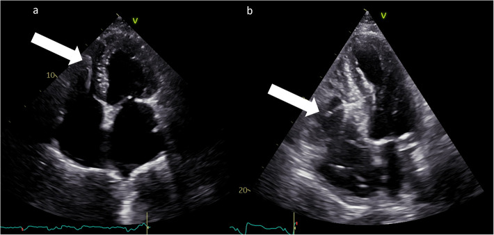
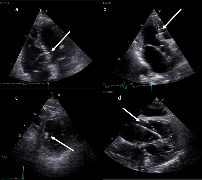
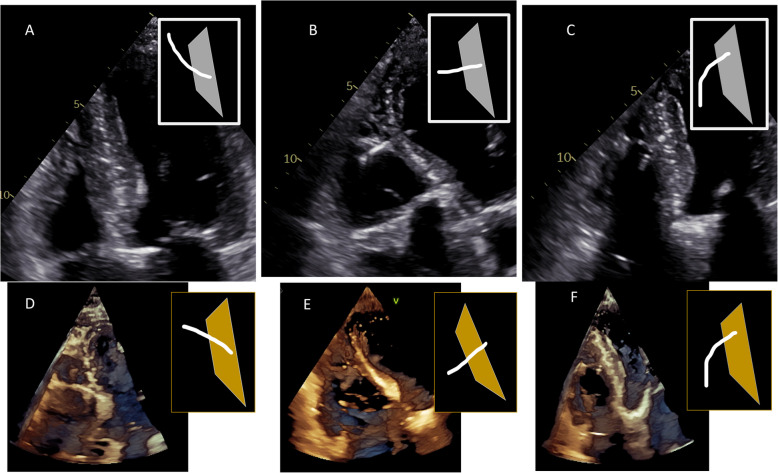
Conduction system pacing (CSP), encompassing His-bundle pacing (HBP) and left bundle branch area pacing (LBBAP), revolutionizes cardiac pacing, allowing a more physiological left ventricular activation than conventional right ventricular (RV) pacing through electrode placed in RV apex, interventricular septum or right ventricular outflow tract. Echocardiography plays a pivotal role in patient assessment, primarily by measuring left ventricular ejection fraction (LVEF) to determine the pacing strategy in alignment with current guidelines. Clinical data, simulations and ongoing trials on CSP explore CSP viability across various LVEF conditions. CSP is supposed to defer pacing-induced cardiomyopathy (PiCM) associated with conventional right ventricular pacing (RVP). This paper aims to review the current literature regarding the use of echocardiography in CSP. Images from our experience in the echocardiographic lab were used throughout this document to show our proposals of imaging in CSP. Echocardiography may help to determine lead localization within the interventricular septum (IVS), customizing pacing to individual anatomy and electromechanical indices (like atro-ventricular delay) and evaluates often-overlooked valvular function, a potential PiCM contributor. Three-dimensional (3-D) echocardiography widens the knowledge of lead localization and valvular dysfunction, as well as dyssynchrony assessment. Dyssynchrony, crucial both to resynchronization per se and physiological stimulation is quantified via echocardiography, especially using speckle-tracking imaging. Baseline LVEF and follow-up observation of CSP effects: early in Global Longitudinal Strain (GLS), afterwards in LV volumes and LVEF may improve the future proper qualification of patients. Limited left atrial (LA) and right atrial (RA) strain assessments hold potential in the CSP qualification and response assessment context. Echocardiography complements other imaging modalities for comprehensive patient evaluation. Echocardiography is integral in the CSP clinical use, from patient selection (by showing subtle changes in myocardial function) to post-procedure follow-up (tricuspid regurgitation, LV and RV function, leads and synchrony assessment). GLS, assessed by speckle tracking imaging and profound 2D and 3D (lead placement, septum morphology and global heart function under CSP) analyses show promise in CSP outcome assessment, though standardization is needed.
期刊介绍:
Cardiovascular Ultrasound is an online journal, publishing peer-reviewed: original research; authoritative reviews; case reports on challenging and/or unusual diagnostic aspects; and expert opinions on new techniques and technologies. We are particularly interested in articles that include relevant images or video files, which provide an additional dimension to published articles and enhance understanding.
As an open access journal, Cardiovascular Ultrasound ensures high visibility for authors in addition to providing an up-to-date and freely available resource for the community. The journal welcomes discussion, and provides a forum for publishing opinion and debate ranging from biology to engineering to clinical echocardiography, with both speed and versatility.
 求助内容:
求助内容: 应助结果提醒方式:


