{"title":"Chemotherapy elevates cell surface PD-L1 and MHC-I expression in apoptotic gastric cancer cells.","authors":"You-Syuan Lou, Xu-Chen Liu, Chih-Cheng Cheng, Yi-Hsuan Yin, Tzu-Cheng Chien, Pei-Ling Hsu, Chu-Wan Lee, Hsin-Hsien Yu, Bor-Chyuan Su","doi":"10.1177/03946320251338662","DOIUrl":null,"url":null,"abstract":"<p><strong>Background: </strong>The programmed cell death-ligand 1 (PD-L1) combined positive score is used as a patient selection tool and predictive factor for anti-programmed cell death-1 (PD-1)/PD-L1 therapy in gastric cancer. However, the expression of PD-L1 and major histocompatibility complex class I (MHC-I) can be affected by conventional treatment approaches.</p><p><strong>Objective: </strong>In this study, we examined the effects of chemotherapy on surface PD-L1 and surface MHC-I expression in living and apoptotic gastric cancer cells. AGS (moderately differentiated) and SNU-1 (poorly differentiated) cells were treated 5-Fluorouracil (5-Fu), cisplatin, mitomycin c (MMC), and FOLFOX (5-Fu, leucovorin, and oxaliplatin).</p><p><strong>Methods: </strong>To quantify the expression levels of surface PD-L1 or surface MHC-I on living and apoptotic cells, the cells were co-stained with annexin V and PD-L1 or MHC-I antibodies. The percentages of single positive (annexin V-negative, PD-L1-positive; annexin V-negative, MHC-I-positive) and double positive (annexin V-positive, PD-L1-positive; annexin V-positive, MHC-I-positive) cells were analyzed by flow cytometry.</p><p><strong>Results: </strong>Every tested chemotherapeutic agent increased the levels of surface PD-L1 and surface MHC-I, although the extents of increase differed in AGS and SNU-1 cells. In AGS cells, 5-Fu caused the largest increases in surface PD-L1 and surface MHC-I. However, 5-Fu caused the weakest increases in surface PD-L1 and surface MHC-I in SNU-1 cells. Notably, chemotherapy-mediated increases in surface PD-L1 and surface MHC-I mostly occurred on apoptotic cells.</p><p><strong>Conclusion: </strong>Our findings reveal that chemotherapy mainly increases surface PD-L1 and surface MHC-I on apoptotic gastric cancer cells.</p>","PeriodicalId":48647,"journal":{"name":"International Journal of Immunopathology and Pharmacology","volume":"39 ","pages":"3946320251338662"},"PeriodicalIF":2.6000,"publicationDate":"2025-01-01","publicationTypes":"Journal Article","fieldsOfStudy":null,"isOpenAccess":false,"openAccessPdf":"https://www.ncbi.nlm.nih.gov/pmc/articles/PMC12085758/pdf/","citationCount":"0","resultStr":null,"platform":"Semanticscholar","paperid":null,"PeriodicalName":"International Journal of Immunopathology and Pharmacology","FirstCategoryId":"3","ListUrlMain":"https://doi.org/10.1177/03946320251338662","RegionNum":3,"RegionCategory":"医学","ArticlePicture":[],"TitleCN":null,"AbstractTextCN":null,"PMCID":null,"EPubDate":"2025/5/17 0:00:00","PubModel":"Epub","JCR":"","JCRName":"","Score":null,"Total":0}
引用次数: 0
Abstract
Background: The programmed cell death-ligand 1 (PD-L1) combined positive score is used as a patient selection tool and predictive factor for anti-programmed cell death-1 (PD-1)/PD-L1 therapy in gastric cancer. However, the expression of PD-L1 and major histocompatibility complex class I (MHC-I) can be affected by conventional treatment approaches.
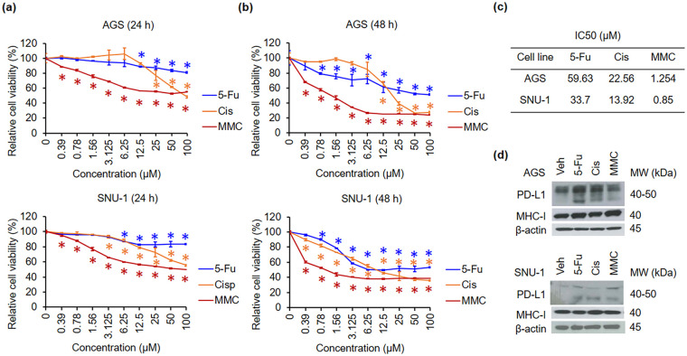
Objective: In this study, we examined the effects of chemotherapy on surface PD-L1 and surface MHC-I expression in living and apoptotic gastric cancer cells. AGS (moderately differentiated) and SNU-1 (poorly differentiated) cells were treated 5-Fluorouracil (5-Fu), cisplatin, mitomycin c (MMC), and FOLFOX (5-Fu, leucovorin, and oxaliplatin).
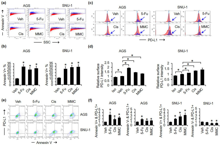
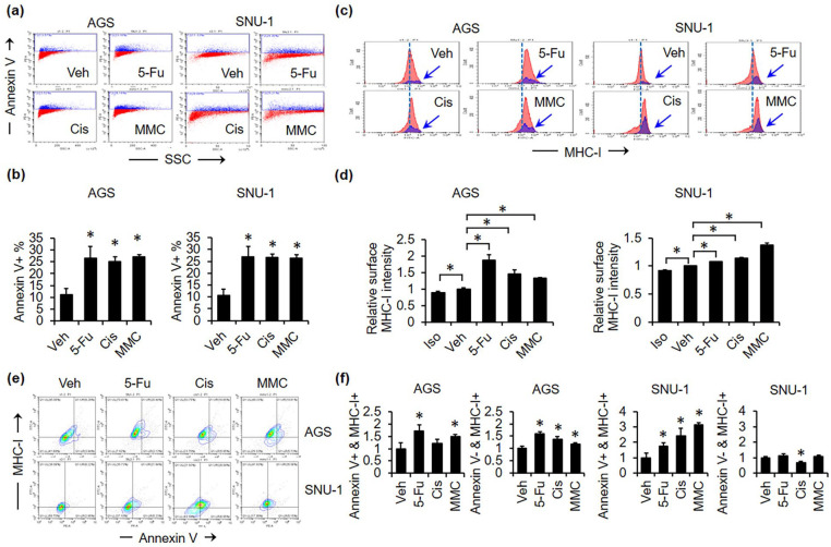
Methods: To quantify the expression levels of surface PD-L1 or surface MHC-I on living and apoptotic cells, the cells were co-stained with annexin V and PD-L1 or MHC-I antibodies. The percentages of single positive (annexin V-negative, PD-L1-positive; annexin V-negative, MHC-I-positive) and double positive (annexin V-positive, PD-L1-positive; annexin V-positive, MHC-I-positive) cells were analyzed by flow cytometry.
Results: Every tested chemotherapeutic agent increased the levels of surface PD-L1 and surface MHC-I, although the extents of increase differed in AGS and SNU-1 cells. In AGS cells, 5-Fu caused the largest increases in surface PD-L1 and surface MHC-I. However, 5-Fu caused the weakest increases in surface PD-L1 and surface MHC-I in SNU-1 cells. Notably, chemotherapy-mediated increases in surface PD-L1 and surface MHC-I mostly occurred on apoptotic cells.
Conclusion: Our findings reveal that chemotherapy mainly increases surface PD-L1 and surface MHC-I on apoptotic gastric cancer cells.
期刊介绍:
International Journal of Immunopathology and Pharmacology is an Open Access peer-reviewed journal publishing original papers describing research in the fields of immunology, pathology and pharmacology. The intention is that the journal should reflect both the experimental and clinical aspects of immunology as well as advances in the understanding of the pathology and pharmacology of the immune system.
 求助内容:
求助内容: 应助结果提醒方式:


