Quantification of perivascular adipose tissue attenuation does not add incremental prognostic value in patients undergoing transcatheter aortic valve implantation.
Carolina Donà, Noemi Pavo, Adriana Vinzens, Pimrapat Gebert, Dietrich Beitzke, Lukas Reider, Nidaa Mikail, Alexia Rossi, Katharina Mascherbauer, Susan Bengs, Achi Haider, Ronny R Buechel, Philipp E Bartko, Christian Loewe, Julia Mascherbauer, Christian Hengstenberg, Georg Goliasch, Max Paul Winter, Catherine Gebhard
{"title":"Quantification of perivascular adipose tissue attenuation does not add incremental prognostic value in patients undergoing transcatheter aortic valve implantation.","authors":"Carolina Donà, Noemi Pavo, Adriana Vinzens, Pimrapat Gebert, Dietrich Beitzke, Lukas Reider, Nidaa Mikail, Alexia Rossi, Katharina Mascherbauer, Susan Bengs, Achi Haider, Ronny R Buechel, Philipp E Bartko, Christian Loewe, Julia Mascherbauer, Christian Hengstenberg, Georg Goliasch, Max Paul Winter, Catherine Gebhard","doi":"10.1093/ehjimp/qyaf047","DOIUrl":null,"url":null,"abstract":"<p><strong>Aims: </strong>Perivascular adipose tissue attenuation (PVAT) has emerged as a novel coronary computed tomography angiography (CCTA)-based biomarker predicting cardiovascular events by capturing inflammation around the coronary arteries. We assessed whether PVAT adds incremental prognostic value in patients undergoing transcatheter aortic valve implantation (TAVI).</p><p><strong>Methods and results: </strong>A total of 510 patients underwent CCTA imaging prior to TAVI between November 2015 and June 2020 at the Medical University of Vienna. PVAT was obtained from CCTA images and was measured around the right coronary artery [PVAT(RCA)] and the aortic valve [PVAT(valve)]. Following application of exclusion criteria, 372 patients [mean age 80.6 ± 6.8 years; 169 (45%) women] were analysed. Over a median follow-up of 3.0 (IQR 2.5-3.6) years, 52 (14%) individuals experienced a major adverse cardiovascular event (MACE, a composite of non-fatal stroke or myocardial infarction, cardiac death, or vascular intervention). Individuals exhibiting elevated PVAT[valve] displayed a heightened surgical risk according to European System for Cardiac Operative Risk Evaluation II, a lower body mass index, reduced left ventricular ejection fraction, prolonged hospitalization following TAVI, and elevated levels of circulating inflammatory markers compared with those in the low PVAT[valve] group (<i>P</i> < 0.05). However, neither PVAT[valve] nor PVAT[RCA] were independently associated with the occurrence of MACE in adjusted multi-variable analyses (PVAT[valve]: sub-distribution hazard ratio [SHR] 1.14, 95% CI:0.63-2.05, <i>P</i> = 0.672); PVAT[RCA]: SHR 1.16 [95% CI: 0.81-1.66], <i>P</i> = 0.417).</p><p><strong>Conclusion: </strong>Measuring PVAT around either the right coronary artery or the aortic valve does not provide additional prognostic value beyond established risk factors for the prediction of MACE in patients undergoing TAVI.</p>","PeriodicalId":94317,"journal":{"name":"European heart journal. Imaging methods and practice","volume":"3 1","pages":"qyaf047"},"PeriodicalIF":0.0000,"publicationDate":"2025-04-18","publicationTypes":"Journal Article","fieldsOfStudy":null,"isOpenAccess":false,"openAccessPdf":"https://www.ncbi.nlm.nih.gov/pmc/articles/PMC12078938/pdf/","citationCount":"0","resultStr":null,"platform":"Semanticscholar","paperid":null,"PeriodicalName":"European heart journal. Imaging methods and practice","FirstCategoryId":"1085","ListUrlMain":"https://doi.org/10.1093/ehjimp/qyaf047","RegionNum":0,"RegionCategory":null,"ArticlePicture":[],"TitleCN":null,"AbstractTextCN":null,"PMCID":null,"EPubDate":"2025/1/1 0:00:00","PubModel":"eCollection","JCR":"","JCRName":"","Score":null,"Total":0}
引用次数: 0
Abstract
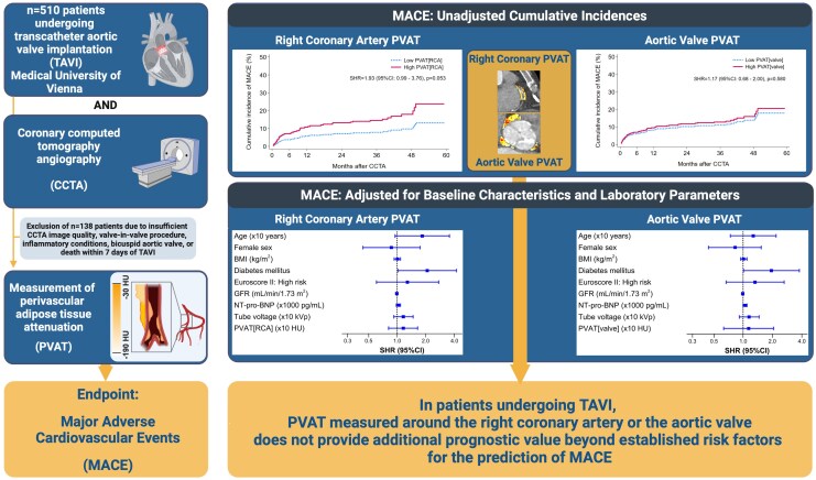
Aims: Perivascular adipose tissue attenuation (PVAT) has emerged as a novel coronary computed tomography angiography (CCTA)-based biomarker predicting cardiovascular events by capturing inflammation around the coronary arteries. We assessed whether PVAT adds incremental prognostic value in patients undergoing transcatheter aortic valve implantation (TAVI).
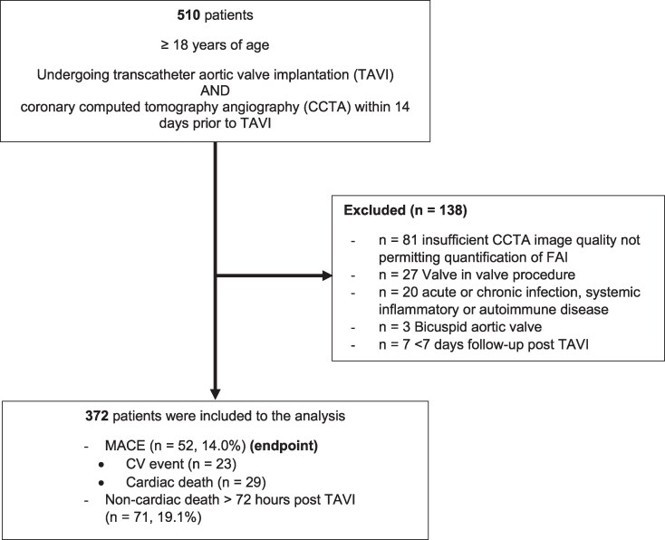
Methods and results: A total of 510 patients underwent CCTA imaging prior to TAVI between November 2015 and June 2020 at the Medical University of Vienna. PVAT was obtained from CCTA images and was measured around the right coronary artery [PVAT(RCA)] and the aortic valve [PVAT(valve)]. Following application of exclusion criteria, 372 patients [mean age 80.6 ± 6.8 years; 169 (45%) women] were analysed. Over a median follow-up of 3.0 (IQR 2.5-3.6) years, 52 (14%) individuals experienced a major adverse cardiovascular event (MACE, a composite of non-fatal stroke or myocardial infarction, cardiac death, or vascular intervention). Individuals exhibiting elevated PVAT[valve] displayed a heightened surgical risk according to European System for Cardiac Operative Risk Evaluation II, a lower body mass index, reduced left ventricular ejection fraction, prolonged hospitalization following TAVI, and elevated levels of circulating inflammatory markers compared with those in the low PVAT[valve] group (P < 0.05). However, neither PVAT[valve] nor PVAT[RCA] were independently associated with the occurrence of MACE in adjusted multi-variable analyses (PVAT[valve]: sub-distribution hazard ratio [SHR] 1.14, 95% CI:0.63-2.05, P = 0.672); PVAT[RCA]: SHR 1.16 [95% CI: 0.81-1.66], P = 0.417).
Conclusion: Measuring PVAT around either the right coronary artery or the aortic valve does not provide additional prognostic value beyond established risk factors for the prediction of MACE in patients undergoing TAVI.

 求助内容:
求助内容: 应助结果提醒方式:


