Ann Mari Svensson, Anna Kistner, Kristina Kairaitis, G Kim Prisk, Catherine Farrow, Terence Amis, Peter D Wagner, Atul Malhotra, Piotr Harbut
{"title":"Quantitative assessment of lung opacities from CT of pulmonary artery imaging data in COVID-19 patients: artificial intelligence versus radiologist.","authors":"Ann Mari Svensson, Anna Kistner, Kristina Kairaitis, G Kim Prisk, Catherine Farrow, Terence Amis, Peter D Wagner, Atul Malhotra, Piotr Harbut","doi":"10.1093/bjro/tzaf008","DOIUrl":null,"url":null,"abstract":"<p><strong>Objectives: </strong>Artificial intelligence (AI) deep learning algorithms trained on non-contrast CT scans effectively detect and quantify acute COVID-19 lung involvement. Our study explored whether radiological contrast affects the accuracy of AI-measured lung opacities, potentially impacting clinical decisions. We compared lung opacity measurements from AI software with visual assessments by radiologists using CT pulmonary angiography (CTPA) images of early-stage COVID-19 patients.</p><p><strong>Methods: </strong>This prospective single-centre study included 18 COVID-19 patients who underwent CTPA due to suspected pulmonary embolism. Patient demographics, clinical data, and 30-day and 90-day mortality were recorded. AI tool (Pulmonary Density Plug-in, AI-Rad Companion Chest CT, SyngoVia; Siemens Healthineers, Forchheim, Germany) was used to estimate the quantity of opacities. Visual quantitative assessments were performed independently by 2 radiologists.</p><p><strong>Results: </strong>There was a positive correlation between radiologist estimations (<i>r</i> <sup>2</sup> = 0.57) and between the AI data and the mean of the radiologists' estimations (<i>r</i> <sup>2</sup> = 0.70). Bland-Altman plot analysis showed a mean bias of +3.06% between radiologists and -1.32% between the mean radiologist vs AI, with no outliers outside 2×SD for respective comparison.</p><p><p>The AI protocol facilitated a quantitative assessment of lung opacities and showed a strong correlation with data obtained from 2 independent radiologists, demonstrating its potential as a complementary tool in clinical practice.</p><p><strong>Conclusion: </strong>In assessing COVID-19 lung opacities in CTPA images, AI tools trained on non-contrast images, provide comparable results to visual assessments by radiologists.</p><p><strong>Advances in knowledge: </strong>The Pulmonary Density Plug-in enables quantitative analysis of lung opacities in COVID-19 patients using contrast-enhanced CT images, potentially streamlining clinical workflows and supporting timely decision-making.</p>","PeriodicalId":72419,"journal":{"name":"BJR open","volume":"7 1","pages":"tzaf008"},"PeriodicalIF":2.1000,"publicationDate":"2025-04-29","publicationTypes":"Journal Article","fieldsOfStudy":null,"isOpenAccess":false,"openAccessPdf":"https://www.ncbi.nlm.nih.gov/pmc/articles/PMC12077292/pdf/","citationCount":"0","resultStr":null,"platform":"Semanticscholar","paperid":null,"PeriodicalName":"BJR open","FirstCategoryId":"1085","ListUrlMain":"https://doi.org/10.1093/bjro/tzaf008","RegionNum":0,"RegionCategory":null,"ArticlePicture":[],"TitleCN":null,"AbstractTextCN":null,"PMCID":null,"EPubDate":"2025/1/1 0:00:00","PubModel":"eCollection","JCR":"","JCRName":"","Score":null,"Total":0}
引用次数: 0
Abstract
Objectives: Artificial intelligence (AI) deep learning algorithms trained on non-contrast CT scans effectively detect and quantify acute COVID-19 lung involvement. Our study explored whether radiological contrast affects the accuracy of AI-measured lung opacities, potentially impacting clinical decisions. We compared lung opacity measurements from AI software with visual assessments by radiologists using CT pulmonary angiography (CTPA) images of early-stage COVID-19 patients.
Methods: This prospective single-centre study included 18 COVID-19 patients who underwent CTPA due to suspected pulmonary embolism. Patient demographics, clinical data, and 30-day and 90-day mortality were recorded. AI tool (Pulmonary Density Plug-in, AI-Rad Companion Chest CT, SyngoVia; Siemens Healthineers, Forchheim, Germany) was used to estimate the quantity of opacities. Visual quantitative assessments were performed independently by 2 radiologists.
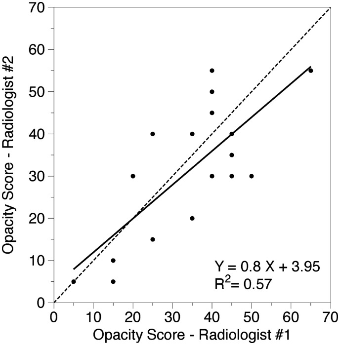
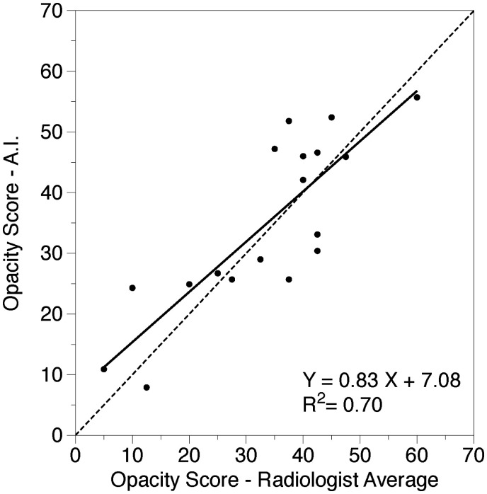
Results: There was a positive correlation between radiologist estimations (r2 = 0.57) and between the AI data and the mean of the radiologists' estimations (r2 = 0.70). Bland-Altman plot analysis showed a mean bias of +3.06% between radiologists and -1.32% between the mean radiologist vs AI, with no outliers outside 2×SD for respective comparison.
The AI protocol facilitated a quantitative assessment of lung opacities and showed a strong correlation with data obtained from 2 independent radiologists, demonstrating its potential as a complementary tool in clinical practice.
Conclusion: In assessing COVID-19 lung opacities in CTPA images, AI tools trained on non-contrast images, provide comparable results to visual assessments by radiologists.
Advances in knowledge: The Pulmonary Density Plug-in enables quantitative analysis of lung opacities in COVID-19 patients using contrast-enhanced CT images, potentially streamlining clinical workflows and supporting timely decision-making.


 求助内容:
求助内容: 应助结果提醒方式:


