{"title":"Expression of HER2 in urothelial carcinoma and its significance.","authors":"Yao Chang, Delong Zhao, Zicheng Wang, Kejia Zhu, Andong Guo, Jishuang Cao, Chenrui Wu, Sentai Ding","doi":"10.1097/CU9.0000000000000249","DOIUrl":null,"url":null,"abstract":"<p><strong>Objectives: </strong>We explored the expression levels and clinical significance of human epidermal growth factor receptor 2 (HER2) in urothelial carcinoma (UC) tissues.</p><p><strong>Materials and methods: </strong>Patient data were reviewed, and 111 paraffin specimens of UC obtained from the Department of Urology, Shandong Provincial Hospital Affiliated to Shandong First Medical University, from 2020 to 2021 were collected. Immunohistochemistry was used to detect HER2 protein expression in all UC tumor tissues. The <i>χ</i> <sup>2</sup> and Fisher exact tests were used to analyze the relationship between HER2 protein expression and clinicopathological data (sex, age, histopathological diagnosis, invasiveness, histopathological grade, maximum tumor diameter, muscle invasion, regional lymph node metastasis, and clinical stage).</p><p><strong>Results: </strong>In this study, 92 cases (82.88%) showed HER2 protein expression, and there was a statistically significant difference in the distribution of HER2 positivity (immunohistochemistry 2+ and 3+) according to the pathological grades of UC (<i>p</i> = 0.021). Human epidermal growth factor receptor 2 positivity was not associated with sex, age, histopathological diagnosis, invasiveness, maximum tumor diameter, muscle invasion, regional lymph node metastasis, or clinical stage (all <i>p</i> < 0.05).</p><p><strong>Conclusions: </strong>Human epidermal growth factor receptor 2 protein is highly expressed in UC, and its expression may be closely related to the high pathological grade of UC.</p>","PeriodicalId":39147,"journal":{"name":"Current Urology","volume":"19 3","pages":"201-207"},"PeriodicalIF":1.3000,"publicationDate":"2025-05-01","publicationTypes":"Journal Article","fieldsOfStudy":null,"isOpenAccess":false,"openAccessPdf":"https://www.ncbi.nlm.nih.gov/pmc/articles/PMC12076403/pdf/","citationCount":"0","resultStr":null,"platform":"Semanticscholar","paperid":null,"PeriodicalName":"Current Urology","FirstCategoryId":"1085","ListUrlMain":"https://doi.org/10.1097/CU9.0000000000000249","RegionNum":4,"RegionCategory":"医学","ArticlePicture":[],"TitleCN":null,"AbstractTextCN":null,"PMCID":null,"EPubDate":"2024/5/27 0:00:00","PubModel":"Epub","JCR":"Q4","JCRName":"UROLOGY & NEPHROLOGY","Score":null,"Total":0}
引用次数: 0
Abstract
Objectives: We explored the expression levels and clinical significance of human epidermal growth factor receptor 2 (HER2) in urothelial carcinoma (UC) tissues.
Materials and methods: Patient data were reviewed, and 111 paraffin specimens of UC obtained from the Department of Urology, Shandong Provincial Hospital Affiliated to Shandong First Medical University, from 2020 to 2021 were collected. Immunohistochemistry was used to detect HER2 protein expression in all UC tumor tissues. The χ2 and Fisher exact tests were used to analyze the relationship between HER2 protein expression and clinicopathological data (sex, age, histopathological diagnosis, invasiveness, histopathological grade, maximum tumor diameter, muscle invasion, regional lymph node metastasis, and clinical stage).
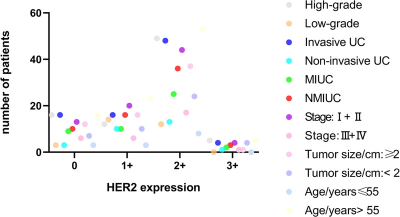
Results: In this study, 92 cases (82.88%) showed HER2 protein expression, and there was a statistically significant difference in the distribution of HER2 positivity (immunohistochemistry 2+ and 3+) according to the pathological grades of UC (p = 0.021). Human epidermal growth factor receptor 2 positivity was not associated with sex, age, histopathological diagnosis, invasiveness, maximum tumor diameter, muscle invasion, regional lymph node metastasis, or clinical stage (all p < 0.05).
Conclusions: Human epidermal growth factor receptor 2 protein is highly expressed in UC, and its expression may be closely related to the high pathological grade of UC.

 求助内容:
求助内容: 应助结果提醒方式:


