Elazir Barbosa Mota Di Puglia, Pedro Augusto Nascimento Daltro, Heron Werner Junior, Miriam Menna Barreto, Flávia Angélica Ferreira Francisco, Sérgio Ferreira Alves Junior, Ivonete Siviero, Claudia Renata S Paio Rezende, Edson Marchiori
{"title":"Ultrasound findings for the diagnosis of biliary atresia in neonates.","authors":"Elazir Barbosa Mota Di Puglia, Pedro Augusto Nascimento Daltro, Heron Werner Junior, Miriam Menna Barreto, Flávia Angélica Ferreira Francisco, Sérgio Ferreira Alves Junior, Ivonete Siviero, Claudia Renata S Paio Rezende, Edson Marchiori","doi":"10.1590/0100-3984.2024.0102","DOIUrl":null,"url":null,"abstract":"<p><strong>Objective: </strong>To investigate and identify the main abdominal ultrasound findings in patients with biliary atresia (BA).</p><p><strong>Materials and methods: </strong>This was a retrospective study of the ultrasound images of 44 patients with neonatal cholestasis. We excluded 18 patients in whom a final diagnosis of BA was not confirmed or who were lost to clinical follow-up. The main ultrasound findings evaluated were gallbladder length and morphology; triangular cord thickness; hepatic artery enlargement; hepatic subcapsular flow; cysts in the porta hepatis; presence of a distinct triangular cord with linear, tubular, or round hypoechoic portions; and polysplenia syndrome.</p><p><strong>Results: </strong>Abnormal gallbladder morphology and triangular cord thickening were the main ultrasound findings in the patients with BA. Gallbladder abnormalities were present in all patients. Hepatic artery enlargement was the third most common finding, present in 19 (73%) patients. Six patients (23%) had subcapsular arterial flow and four (15%) had cysts in the porta hepatis. Hypoechoic or cystic portions of the triangular cord were present in three patients (11%), and we found that BA was accompanied by polysplenia syndrome in three patients (11%).</p><p><strong>Conclusion: </strong>Ultrasound is the examination of greatest diagnostic relevance in the investigation of cholestasis in newborns and infants; it enables the establishment of BA suspicion and the indication for laparotomy with intraoperative cholangiography.</p>","PeriodicalId":20842,"journal":{"name":"Radiologia Brasileira","volume":"58 ","pages":"e20240102"},"PeriodicalIF":0.0000,"publicationDate":"2025-04-25","publicationTypes":"Journal Article","fieldsOfStudy":null,"isOpenAccess":false,"openAccessPdf":"https://www.ncbi.nlm.nih.gov/pmc/articles/PMC12076785/pdf/","citationCount":"0","resultStr":null,"platform":"Semanticscholar","paperid":null,"PeriodicalName":"Radiologia Brasileira","FirstCategoryId":"1085","ListUrlMain":"https://doi.org/10.1590/0100-3984.2024.0102","RegionNum":0,"RegionCategory":null,"ArticlePicture":[],"TitleCN":null,"AbstractTextCN":null,"PMCID":null,"EPubDate":"2025/1/1 0:00:00","PubModel":"eCollection","JCR":"Q3","JCRName":"Medicine","Score":null,"Total":0}
引用次数: 0
Abstract
Objective: To investigate and identify the main abdominal ultrasound findings in patients with biliary atresia (BA).
Materials and methods: This was a retrospective study of the ultrasound images of 44 patients with neonatal cholestasis. We excluded 18 patients in whom a final diagnosis of BA was not confirmed or who were lost to clinical follow-up. The main ultrasound findings evaluated were gallbladder length and morphology; triangular cord thickness; hepatic artery enlargement; hepatic subcapsular flow; cysts in the porta hepatis; presence of a distinct triangular cord with linear, tubular, or round hypoechoic portions; and polysplenia syndrome.
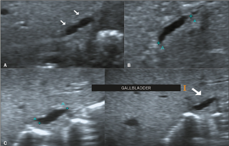
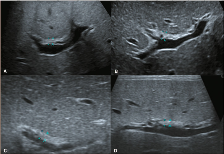
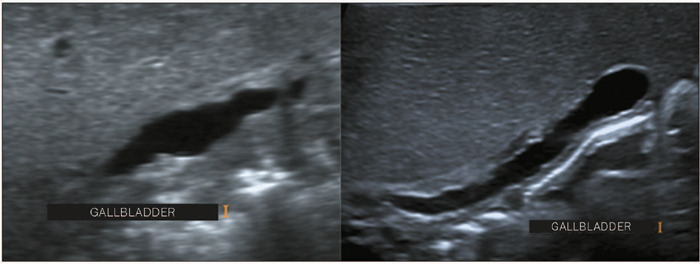
Results: Abnormal gallbladder morphology and triangular cord thickening were the main ultrasound findings in the patients with BA. Gallbladder abnormalities were present in all patients. Hepatic artery enlargement was the third most common finding, present in 19 (73%) patients. Six patients (23%) had subcapsular arterial flow and four (15%) had cysts in the porta hepatis. Hypoechoic or cystic portions of the triangular cord were present in three patients (11%), and we found that BA was accompanied by polysplenia syndrome in three patients (11%).
Conclusion: Ultrasound is the examination of greatest diagnostic relevance in the investigation of cholestasis in newborns and infants; it enables the establishment of BA suspicion and the indication for laparotomy with intraoperative cholangiography.

 求助内容:
求助内容: 应助结果提醒方式:


