{"title":"Integrative Analysis of <sup>18</sup>F-FDG PET Radiomics and mRNA Expression in Recurrent/Metastatic Oral Squamous Cell Carcinoma: A Cross-Sectional Study.","authors":"Mai Kim, Wenchao Gu, Reika Kawabata- Iwakawa, Shinichiro Kina, Takahito Nakajima, Tetsuya Higuchi, Masaru Ogawa, Keisuke Suzuki, Yoshito Tsushima, Satoshi Yokoo","doi":"10.1007/s11307-025-02012-5","DOIUrl":null,"url":null,"abstract":"<p><strong>Background: </strong>This study explored the relationship between mRNA expression profiles obtained through next-generation sequencing (NGS) and <sup>18</sup>F-fluorodeoxyglucose positron emission tomography (<sup>18</sup>F-FDG PET) texture analysis in patients with treatment-resistant oral squamous cell carcinoma (OSCC) who were treated with molecular-targeted drugs. We analyzed the correlation between <sup>18</sup>F-FDG PET texture features and NGS data in a small cohort of five patients with recurrent or metastatic OSCC who received molecular-targeted drugs after surgery. Patients were categorized into two groups based on treatment response: responders (n = 3) and non-responders (n = 2). To validate our findings, we examined transcriptomic data from 292 OSCC patients in The Cancer Genome Atlas (TCGA) database.</p><p><strong>Results: </strong>The gene ankyrin repeat and SOCS box containing two (ASB2) was significantly overexpressed in non-responders and strongly correlated with specific PET radiomic features, including GLRLM_GLNU, GLRLM_RLNU, and GLZLM_GLNU (p < 0.05). High ASB2 expression was also associated with poor prognosis in OSCC patients (p < 0.05) and decreased overall survival, as shown by Kaplan-Meier analysis of the TCGA database (p = 0.017).</p><p><strong>Conclusions: </strong>Integrating ASB2 expression data with <sup>18</sup>F-FDG PET texture features could potentially improve the prediction of treatment outcomes in treatment-resistant OSCC patients undergoing molecular-targeted therapy.</p>","PeriodicalId":18760,"journal":{"name":"Molecular Imaging and Biology","volume":" ","pages":"421-430"},"PeriodicalIF":2.5000,"publicationDate":"2025-06-01","publicationTypes":"Journal Article","fieldsOfStudy":null,"isOpenAccess":false,"openAccessPdf":"https://www.ncbi.nlm.nih.gov/pmc/articles/PMC12162752/pdf/","citationCount":"0","resultStr":null,"platform":"Semanticscholar","paperid":null,"PeriodicalName":"Molecular Imaging and Biology","FirstCategoryId":"3","ListUrlMain":"https://doi.org/10.1007/s11307-025-02012-5","RegionNum":4,"RegionCategory":"医学","ArticlePicture":[],"TitleCN":null,"AbstractTextCN":null,"PMCID":null,"EPubDate":"2025/5/14 0:00:00","PubModel":"Epub","JCR":"Q2","JCRName":"RADIOLOGY, NUCLEAR MEDICINE & MEDICAL IMAGING","Score":null,"Total":0}
引用次数: 0
Abstract
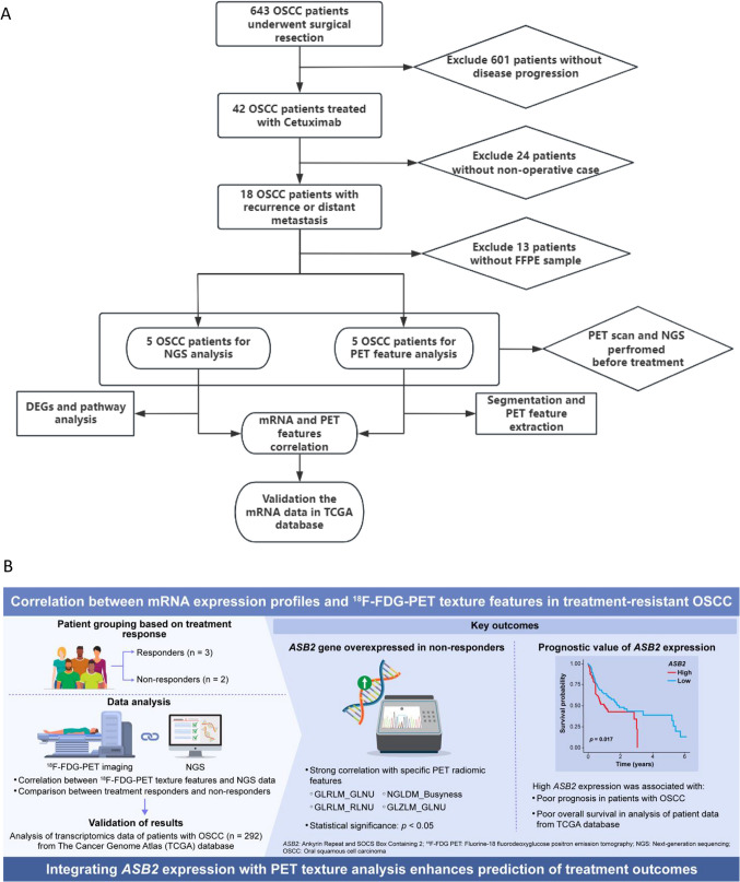
Background: This study explored the relationship between mRNA expression profiles obtained through next-generation sequencing (NGS) and 18F-fluorodeoxyglucose positron emission tomography (18F-FDG PET) texture analysis in patients with treatment-resistant oral squamous cell carcinoma (OSCC) who were treated with molecular-targeted drugs. We analyzed the correlation between 18F-FDG PET texture features and NGS data in a small cohort of five patients with recurrent or metastatic OSCC who received molecular-targeted drugs after surgery. Patients were categorized into two groups based on treatment response: responders (n = 3) and non-responders (n = 2). To validate our findings, we examined transcriptomic data from 292 OSCC patients in The Cancer Genome Atlas (TCGA) database.
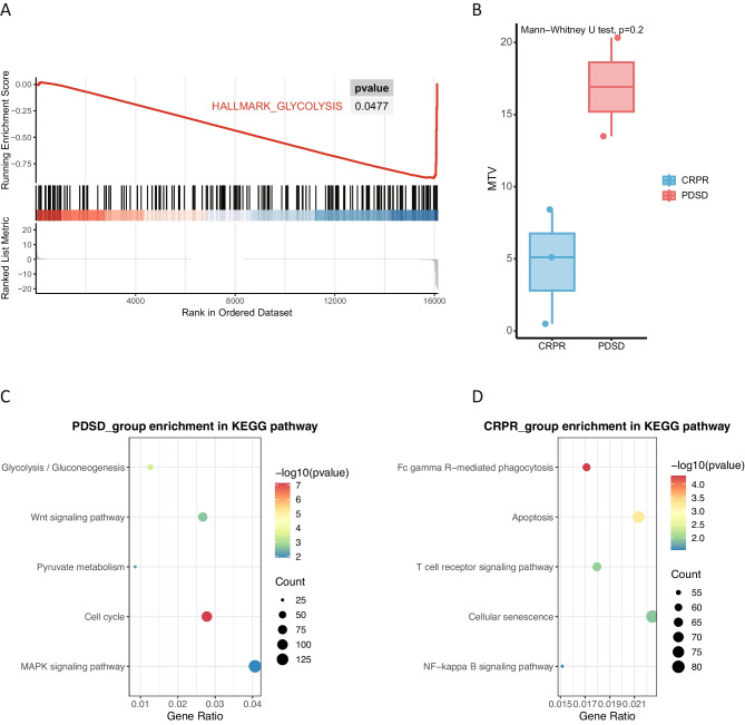
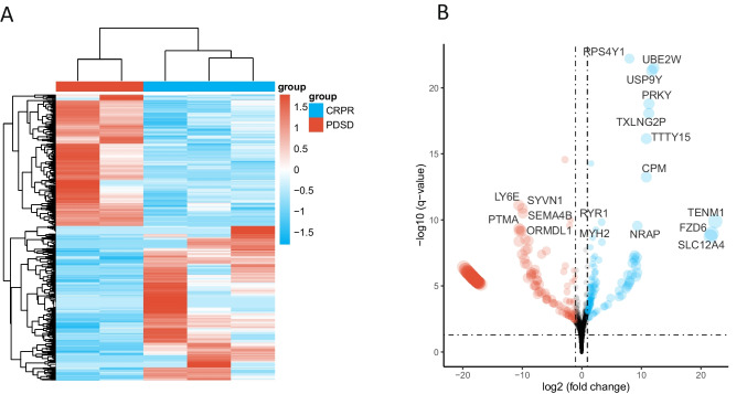
Results: The gene ankyrin repeat and SOCS box containing two (ASB2) was significantly overexpressed in non-responders and strongly correlated with specific PET radiomic features, including GLRLM_GLNU, GLRLM_RLNU, and GLZLM_GLNU (p < 0.05). High ASB2 expression was also associated with poor prognosis in OSCC patients (p < 0.05) and decreased overall survival, as shown by Kaplan-Meier analysis of the TCGA database (p = 0.017).
Conclusions: Integrating ASB2 expression data with 18F-FDG PET texture features could potentially improve the prediction of treatment outcomes in treatment-resistant OSCC patients undergoing molecular-targeted therapy.
期刊介绍:
Molecular Imaging and Biology (MIB) invites original contributions (research articles, review articles, commentaries, etc.) on the utilization of molecular imaging (i.e., nuclear imaging, optical imaging, autoradiography and pathology, MRI, MPI, ultrasound imaging, radiomics/genomics etc.) to investigate questions related to biology and health. The objective of MIB is to provide a forum to the discovery of molecular mechanisms of disease through the use of imaging techniques. We aim to investigate the biological nature of disease in patients and establish new molecular imaging diagnostic and therapy procedures.
Some areas that are covered are:
Preclinical and clinical imaging of macromolecular targets (e.g., genes, receptors, enzymes) involved in significant biological processes.
The design, characterization, and study of new molecular imaging probes and contrast agents for the functional interrogation of macromolecular targets.
Development and evaluation of imaging systems including instrumentation, image reconstruction algorithms, image analysis, and display.
Development of molecular assay approaches leading to quantification of the biological information obtained in molecular imaging.
Study of in vivo animal models of disease for the development of new molecular diagnostics and therapeutics.
Extension of in vitro and in vivo discoveries using disease models, into well designed clinical research investigations.
Clinical molecular imaging involving clinical investigations, clinical trials and medical management or cost-effectiveness studies.

 求助内容:
求助内容: 应助结果提醒方式:


