[18 F]-Fluoroestradiol PET (FES-PET) and [18 F] Flurodeoxyglucose PET (FDG-PET) Imaging May Aid in Managing Therapy in Patients with Metastatic Lobular Breast Cancer.
IF 2.5 4区 医学Q2 RADIOLOGY, NUCLEAR MEDICINE & MEDICAL IMAGING
Poorni M Manohar, Lanell M Peterson, Isaac C Jenkins, Qian Vicky Wu, Brenda F Kurland, Alena Novakova-Jiresova, Mark Muzi, Delphine L Chen, Jennifer M Specht, Suzanne Dintzis, Paul E Kinahan, David A Mankoff, Hannah M Linden
{"title":"[18 F]-Fluoroestradiol PET (FES-PET) and [18 F] Flurodeoxyglucose PET (FDG-PET) Imaging May Aid in Managing Therapy in Patients with Metastatic Lobular Breast Cancer.","authors":"Poorni M Manohar, Lanell M Peterson, Isaac C Jenkins, Qian Vicky Wu, Brenda F Kurland, Alena Novakova-Jiresova, Mark Muzi, Delphine L Chen, Jennifer M Specht, Suzanne Dintzis, Paul E Kinahan, David A Mankoff, Hannah M Linden","doi":"10.1007/s11307-025-02015-2","DOIUrl":null,"url":null,"abstract":"<p><strong>Aim: </strong>This study examines the combination of FES-PET and FDG-PET as complementary imaging for detection of metastatic ILC.</p><p><strong>Methods: </strong>We retrospectively evaluated FES and FDG uptake in patients with metastatic ILC from an estrogen receptor (ER) positive primary tumor. We classified lesions into three categories (FES high/FDG low, FES high/FDG high, FES low/FDG low) using SUVmax cut-off values of 1.5 for FES and 5.0 for FDG. Qualitative evaluation included examination of the difference in the extent of disease between FES and FDG.</p><p><strong>Results: </strong>Of the 38 patients, 82% had FES uptake in all tumor sites identified by FDG, with 18% lacking FES uptake in at least one lesion. Mean (range) SUVmax for FES and FDG was 4.0 (0.67-10.6) and 4.6 (1.3-12.5), respectively. The majority of ILC patients (25/38), had lesions with FES high/FDG low uptake, consistent with the strongly ER + indolent nature of ILC. Patients with disease classified as FES high/FDG low had longer median overall survival (OS) (3.2 years) and progression-free survival (PFS) (1.5 years) than FES high/FDG high (OS = 2.1 years and PFS = 0.46 years). The median overall OS for all patients was 3.0 years (95% CI 2.5, 4.8) and PFS of 1.3 years (95% CI 0.6, 2.5). 8 patients (21%) had qualitatively more extensive disease by FES-PET.</p><p><strong>Conclusions: </strong>Our findings suggest that both FES-PET and FDG-PET can identify metastatic ILC and be useful in detecting the pattern and extent of disease. The imaging combination provides additional information for prognosis and clinical decision making, balancing suitability for endocrine therapy and aggressiveness/indolence of disease.</p>","PeriodicalId":18760,"journal":{"name":"Molecular Imaging and Biology","volume":" ","pages":"410-420"},"PeriodicalIF":2.5000,"publicationDate":"2025-06-01","publicationTypes":"Journal Article","fieldsOfStudy":null,"isOpenAccess":false,"openAccessPdf":"https://www.ncbi.nlm.nih.gov/pmc/articles/PMC12162187/pdf/","citationCount":"0","resultStr":null,"platform":"Semanticscholar","paperid":null,"PeriodicalName":"Molecular Imaging and Biology","FirstCategoryId":"3","ListUrlMain":"https://doi.org/10.1007/s11307-025-02015-2","RegionNum":4,"RegionCategory":"医学","ArticlePicture":[],"TitleCN":null,"AbstractTextCN":null,"PMCID":null,"EPubDate":"2025/5/14 0:00:00","PubModel":"Epub","JCR":"Q2","JCRName":"RADIOLOGY, NUCLEAR MEDICINE & MEDICAL IMAGING","Score":null,"Total":0}
引用次数: 0
Abstract
Aim: This study examines the combination of FES-PET and FDG-PET as complementary imaging for detection of metastatic ILC.
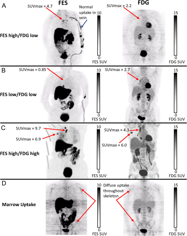
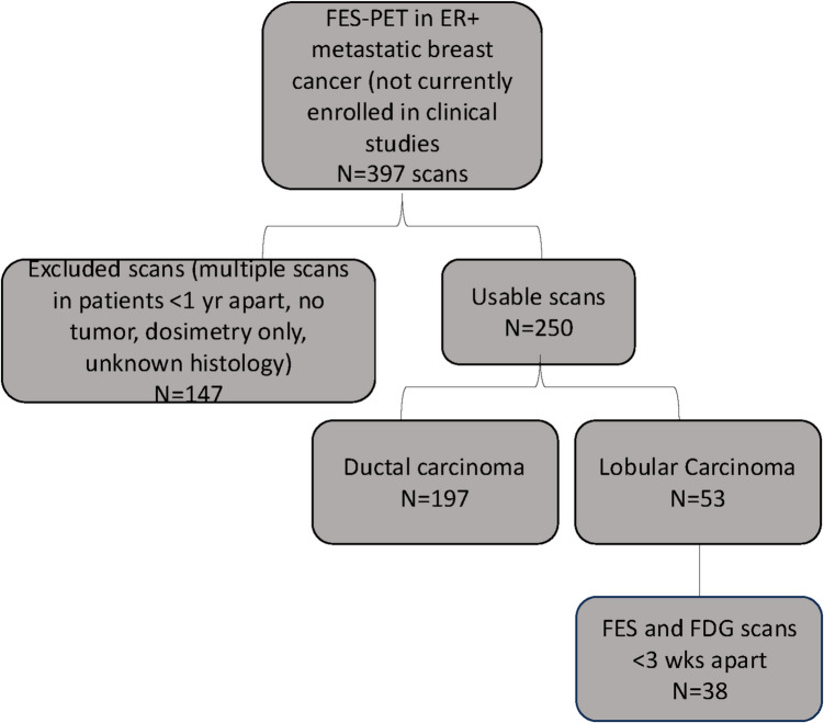
Methods: We retrospectively evaluated FES and FDG uptake in patients with metastatic ILC from an estrogen receptor (ER) positive primary tumor. We classified lesions into three categories (FES high/FDG low, FES high/FDG high, FES low/FDG low) using SUVmax cut-off values of 1.5 for FES and 5.0 for FDG. Qualitative evaluation included examination of the difference in the extent of disease between FES and FDG.
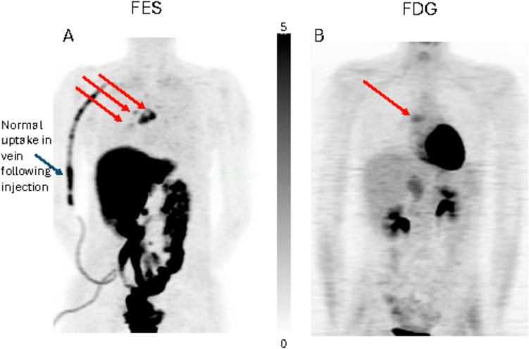
Results: Of the 38 patients, 82% had FES uptake in all tumor sites identified by FDG, with 18% lacking FES uptake in at least one lesion. Mean (range) SUVmax for FES and FDG was 4.0 (0.67-10.6) and 4.6 (1.3-12.5), respectively. The majority of ILC patients (25/38), had lesions with FES high/FDG low uptake, consistent with the strongly ER + indolent nature of ILC. Patients with disease classified as FES high/FDG low had longer median overall survival (OS) (3.2 years) and progression-free survival (PFS) (1.5 years) than FES high/FDG high (OS = 2.1 years and PFS = 0.46 years). The median overall OS for all patients was 3.0 years (95% CI 2.5, 4.8) and PFS of 1.3 years (95% CI 0.6, 2.5). 8 patients (21%) had qualitatively more extensive disease by FES-PET.
Conclusions: Our findings suggest that both FES-PET and FDG-PET can identify metastatic ILC and be useful in detecting the pattern and extent of disease. The imaging combination provides additional information for prognosis and clinical decision making, balancing suitability for endocrine therapy and aggressiveness/indolence of disease.
期刊介绍:
Molecular Imaging and Biology (MIB) invites original contributions (research articles, review articles, commentaries, etc.) on the utilization of molecular imaging (i.e., nuclear imaging, optical imaging, autoradiography and pathology, MRI, MPI, ultrasound imaging, radiomics/genomics etc.) to investigate questions related to biology and health. The objective of MIB is to provide a forum to the discovery of molecular mechanisms of disease through the use of imaging techniques. We aim to investigate the biological nature of disease in patients and establish new molecular imaging diagnostic and therapy procedures.
Some areas that are covered are:
Preclinical and clinical imaging of macromolecular targets (e.g., genes, receptors, enzymes) involved in significant biological processes.
The design, characterization, and study of new molecular imaging probes and contrast agents for the functional interrogation of macromolecular targets.
Development and evaluation of imaging systems including instrumentation, image reconstruction algorithms, image analysis, and display.
Development of molecular assay approaches leading to quantification of the biological information obtained in molecular imaging.
Study of in vivo animal models of disease for the development of new molecular diagnostics and therapeutics.
Extension of in vitro and in vivo discoveries using disease models, into well designed clinical research investigations.
Clinical molecular imaging involving clinical investigations, clinical trials and medical management or cost-effectiveness studies.

 求助内容:
求助内容: 应助结果提醒方式:


