{"title":"White matter and cortical gray matter microstructural alterations in migraine: a NODDI and DTI analysis.","authors":"Zhilei Li, Yanliang Mei, Lei Wang, Tianhua Fan, Cheng Peng, Kaibo Zhang, Shouyi Wu, Tong Chen, Zhenchang Zhang, Binbin Sui, Yonggang Wang, Xueying Yu","doi":"10.1186/s10194-025-02059-3","DOIUrl":null,"url":null,"abstract":"<p><strong>Background: </strong>The pathophysiological mechanism of migraine remains elusive, thereby impeding the effective treatment of the disease. Current neuroimaging research focuses on changes in brain functional connectivity, functional networks, and macrostructural alterations, which reflect abnormal neuronal function during the disease process. The plasticity changes in neuronal structures and neurotransmitter system dysregulations potentially play a crucial role in migraine onset and chronicity of migraine. This study utilizes multimodal neuroimaging techniques to investigate the microstructural and neurotransmitter alterations in migraine and provides new insights into its pathological mechanisms and therapeutic method.</p><p><strong>Methods: </strong>Microstructural alterations in both white matter (WM) and cortical gray matter (GM) were evaluated among 40 chronic migraine (CM) patients, 35 episodic migraine (EM) patients, and 45 healthy controls (HCs) using Diffusion Tensor Imaging (DTI) and Neurite Orientation Dispersion and Density Imaging (NODDI) models. Tract-based spatial statistics (TBSS) and Surface-based analysis (SBA) were performed to compare groupwise differences in white and gray matter microstructure, respectively. Furthermore, the cross-modal toolbox JuSpace was used to analyze the correlation between cortical gray matter neurite alterations and neurotransmitter.</p><p><strong>Results: </strong>In the WM, compared to HC, a decrease in neurite density index (NDI) was identified in the migraine group, and both NDI and fractional anisotropy (FA) were decreased in the CM group. No significant alterations were observed in the EM group. An increase in radial diffusivity (RD) was found in the CM group compared to the EM group. In the cortical GM, compared to HC, the migraine group had fewer neurites in the right insula and temporal pole cortex, and the CM group showed a reduction in neurites in the right middle temporal and fusiform cortex. The cortical GM of neurite damage was negatively correlated with neurotransmitters in migraine and CM. There was no correlation between NODDI and DTI metrics of these brain regions and clinical data after the Bonferroni correction.</p><p><strong>Conclusion: </strong>Our findings indicated that neurite loss was detected in both WM and cortical GM of migraineurs. As the migraine progresses into chronicity, the axonal damage may become more pronounced. The neurite damage of cortical GM was negatively related to neurotransmitters.</p>","PeriodicalId":16013,"journal":{"name":"Journal of Headache and Pain","volume":"26 1","pages":"115"},"PeriodicalIF":7.9000,"publicationDate":"2025-05-14","publicationTypes":"Journal Article","fieldsOfStudy":null,"isOpenAccess":false,"openAccessPdf":"https://www.ncbi.nlm.nih.gov/pmc/articles/PMC12076971/pdf/","citationCount":"0","resultStr":null,"platform":"Semanticscholar","paperid":null,"PeriodicalName":"Journal of Headache and Pain","FirstCategoryId":"3","ListUrlMain":"https://doi.org/10.1186/s10194-025-02059-3","RegionNum":1,"RegionCategory":"医学","ArticlePicture":[],"TitleCN":null,"AbstractTextCN":null,"PMCID":null,"EPubDate":"","PubModel":"","JCR":"Q1","JCRName":"CLINICAL NEUROLOGY","Score":null,"Total":0}
引用次数: 0
Abstract
Background: The pathophysiological mechanism of migraine remains elusive, thereby impeding the effective treatment of the disease. Current neuroimaging research focuses on changes in brain functional connectivity, functional networks, and macrostructural alterations, which reflect abnormal neuronal function during the disease process. The plasticity changes in neuronal structures and neurotransmitter system dysregulations potentially play a crucial role in migraine onset and chronicity of migraine. This study utilizes multimodal neuroimaging techniques to investigate the microstructural and neurotransmitter alterations in migraine and provides new insights into its pathological mechanisms and therapeutic method.
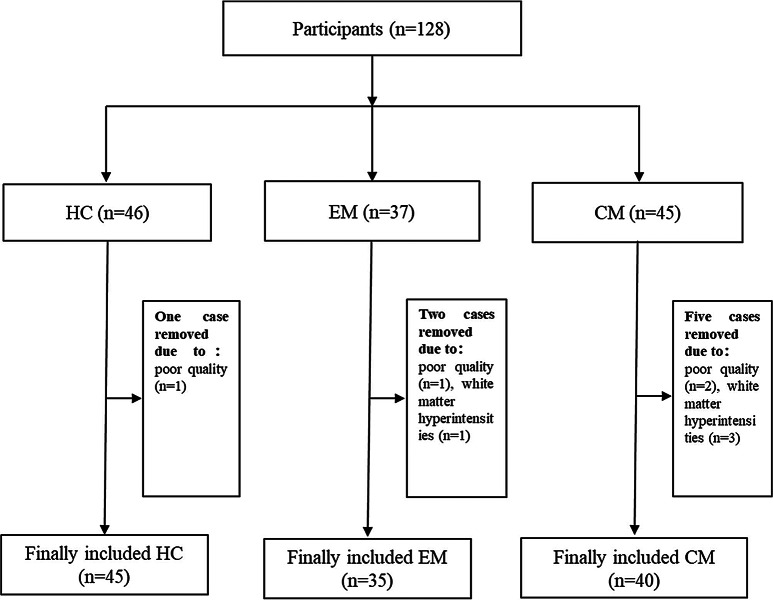
Methods: Microstructural alterations in both white matter (WM) and cortical gray matter (GM) were evaluated among 40 chronic migraine (CM) patients, 35 episodic migraine (EM) patients, and 45 healthy controls (HCs) using Diffusion Tensor Imaging (DTI) and Neurite Orientation Dispersion and Density Imaging (NODDI) models. Tract-based spatial statistics (TBSS) and Surface-based analysis (SBA) were performed to compare groupwise differences in white and gray matter microstructure, respectively. Furthermore, the cross-modal toolbox JuSpace was used to analyze the correlation between cortical gray matter neurite alterations and neurotransmitter.
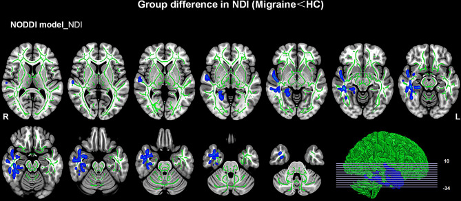
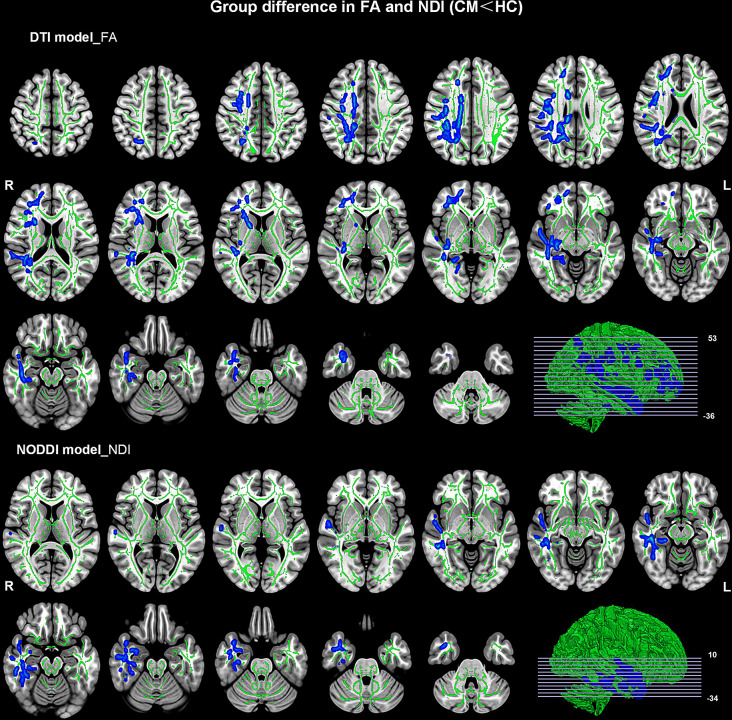
Results: In the WM, compared to HC, a decrease in neurite density index (NDI) was identified in the migraine group, and both NDI and fractional anisotropy (FA) were decreased in the CM group. No significant alterations were observed in the EM group. An increase in radial diffusivity (RD) was found in the CM group compared to the EM group. In the cortical GM, compared to HC, the migraine group had fewer neurites in the right insula and temporal pole cortex, and the CM group showed a reduction in neurites in the right middle temporal and fusiform cortex. The cortical GM of neurite damage was negatively correlated with neurotransmitters in migraine and CM. There was no correlation between NODDI and DTI metrics of these brain regions and clinical data after the Bonferroni correction.
Conclusion: Our findings indicated that neurite loss was detected in both WM and cortical GM of migraineurs. As the migraine progresses into chronicity, the axonal damage may become more pronounced. The neurite damage of cortical GM was negatively related to neurotransmitters.
期刊介绍:
The Journal of Headache and Pain, a peer-reviewed open-access journal published under the BMC brand, a part of Springer Nature, is dedicated to researchers engaged in all facets of headache and related pain syndromes. It encompasses epidemiology, public health, basic science, translational medicine, clinical trials, and real-world data.
With a multidisciplinary approach, The Journal of Headache and Pain addresses headache medicine and related pain syndromes across all medical disciplines. It particularly encourages submissions in clinical, translational, and basic science fields, focusing on pain management, genetics, neurology, and internal medicine. The journal publishes research articles, reviews, letters to the Editor, as well as consensus articles and guidelines, aimed at promoting best practices in managing patients with headaches and related pain.

 求助内容:
求助内容: 应助结果提醒方式:


