David Michaël de Jong, Daniëlle Roosterman, Marco J Bruno, Lydi M J W van Driel, Wim J Lammers
{"title":"Interobserver variability in lymph node evaluation with endoscopic ultrasonography in cholangiocarcinoma.","authors":"David Michaël de Jong, Daniëlle Roosterman, Marco J Bruno, Lydi M J W van Driel, Wim J Lammers","doi":"10.1055/a-2577-5449","DOIUrl":null,"url":null,"abstract":"<p><strong>Background and study aims: </strong>Accurate preoperative lymph node (LN) assessment is crucial for patients with intrahepatic cholangiocarcinoma (iCCA) and perihilar cholangiocarcinoma (pCCA) because presence of LN metastases significantly reduces survival rates and can contraindicate surgical resection. Endoscopic ultrasound (EUS) provides a reliable method for LN assessment with the advantage of enabling tissue acquisition for pathological confirmation. This study aimed to assess interobserver agreement among endosonographers in evaluating LN characteristics in patients with iCCA and pCCA.</p><p><strong>Methods: </strong>A cross-sectional survey study was conducted among 24 endosonographers. Participants reviewed 42 EUS images from iCCA and pCCA patients, classifying LNs based on six characteristics (demarcation, shape, echogenicity, homogeneity, suspiciousness, and need to retrieve tissue). Interobserver agreement was determined using Light's kappa statistics. Accuracy, sensitivity, and specificity in identifying malignant LNs were calculated.</p><p><strong>Results: </strong>Overall kappa values indicated moderate to fair agreement on LN characteristics, with Kappa values of 0.24 for demarcation, 0.45 for shape, 0.38 for echogenicity, 0.52 for homogeneity, and 0.36 for suspiciousness. Overall accuracy of endosonographers in correctly identifying malignant LNs was 62%, with individual accuracy ranging from 44 to 75%. Sensitivity was 60% (range: 29%-90%) and specificity was 64% (range: 28%-89%).</p><p><strong>Conclusions: </strong>Endosonographic assessment of LN morphology and characterization demonstrates considerable variability among endosonographers. Thus, there is a clear need for standardization in preoperative LN evaluation, including establishing consensus about when to perform tissue acquisition, based on objective criteria such as short-axis diameter. Further research is required to refine and optimize these guidelines.</p>","PeriodicalId":11671,"journal":{"name":"Endoscopy International Open","volume":"13 ","pages":"a25775449"},"PeriodicalIF":2.3000,"publicationDate":"2025-05-12","publicationTypes":"Journal Article","fieldsOfStudy":null,"isOpenAccess":false,"openAccessPdf":"https://www.ncbi.nlm.nih.gov/pmc/articles/PMC12080517/pdf/","citationCount":"0","resultStr":null,"platform":"Semanticscholar","paperid":null,"PeriodicalName":"Endoscopy International Open","FirstCategoryId":"1085","ListUrlMain":"https://doi.org/10.1055/a-2577-5449","RegionNum":0,"RegionCategory":null,"ArticlePicture":[],"TitleCN":null,"AbstractTextCN":null,"PMCID":null,"EPubDate":"2025/1/1 0:00:00","PubModel":"eCollection","JCR":"Q3","JCRName":"GASTROENTEROLOGY & HEPATOLOGY","Score":null,"Total":0}
引用次数: 0
Abstract
Background and study aims: Accurate preoperative lymph node (LN) assessment is crucial for patients with intrahepatic cholangiocarcinoma (iCCA) and perihilar cholangiocarcinoma (pCCA) because presence of LN metastases significantly reduces survival rates and can contraindicate surgical resection. Endoscopic ultrasound (EUS) provides a reliable method for LN assessment with the advantage of enabling tissue acquisition for pathological confirmation. This study aimed to assess interobserver agreement among endosonographers in evaluating LN characteristics in patients with iCCA and pCCA.
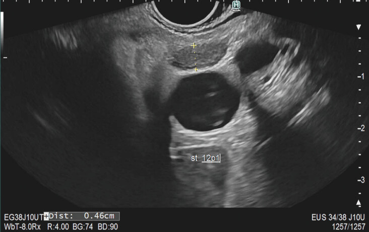
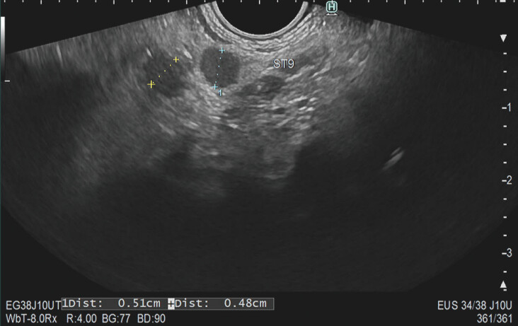
Methods: A cross-sectional survey study was conducted among 24 endosonographers. Participants reviewed 42 EUS images from iCCA and pCCA patients, classifying LNs based on six characteristics (demarcation, shape, echogenicity, homogeneity, suspiciousness, and need to retrieve tissue). Interobserver agreement was determined using Light's kappa statistics. Accuracy, sensitivity, and specificity in identifying malignant LNs were calculated.
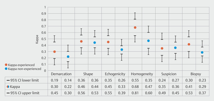
Results: Overall kappa values indicated moderate to fair agreement on LN characteristics, with Kappa values of 0.24 for demarcation, 0.45 for shape, 0.38 for echogenicity, 0.52 for homogeneity, and 0.36 for suspiciousness. Overall accuracy of endosonographers in correctly identifying malignant LNs was 62%, with individual accuracy ranging from 44 to 75%. Sensitivity was 60% (range: 29%-90%) and specificity was 64% (range: 28%-89%).
Conclusions: Endosonographic assessment of LN morphology and characterization demonstrates considerable variability among endosonographers. Thus, there is a clear need for standardization in preoperative LN evaluation, including establishing consensus about when to perform tissue acquisition, based on objective criteria such as short-axis diameter. Further research is required to refine and optimize these guidelines.

 求助内容:
求助内容: 应助结果提醒方式:


